Esophageal varices and portal hypertension
Board exam relevance: in 7 of 105 exam reports · rank 53- Synonyms
- portal hypertension, esophageal varices, varices in the gullet, variceal bleeding, gastric varices, caput medusae
- Specialty
- Internal medicine · Liver & biliary tract
- Images
- Clinical 1 · CT 2 · Ultrasound 1
- Last updated
- 10/2026 · Dr. Pascal Bafteh
Contents
Images (4)
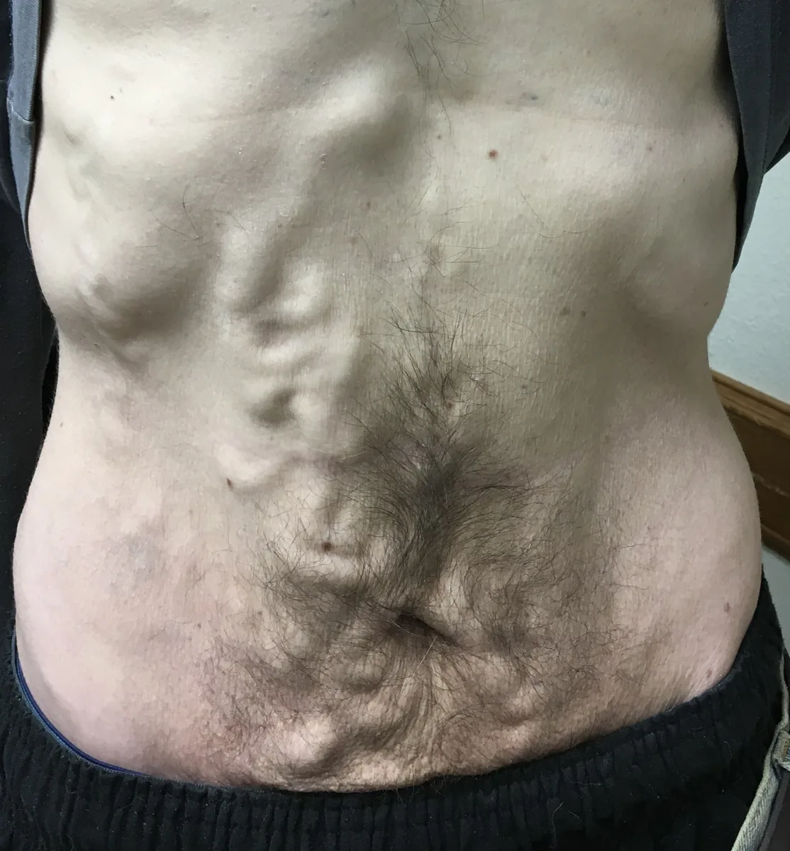
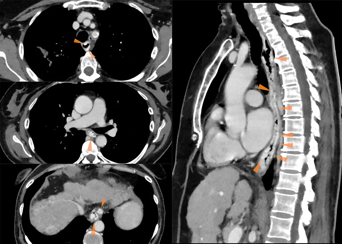 CT
CT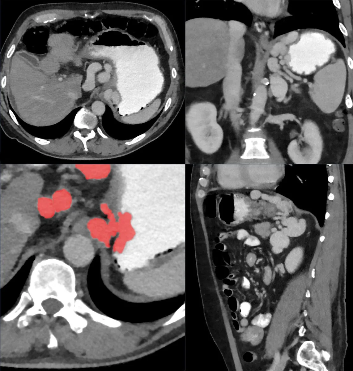 CT
CT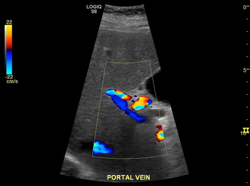 Ultrasound
Definition
Portal hypertension is raised pressure in the portal venous system. Normal portal pressure is 5–10 mmHg, and the gradient to the inferior vena cava is below about 5 mmHg.
Esophageal varices are dilated, tortuous submucosal veins in the distal esophagus, and fundal varices are the corresponding vessels in the gastric fundus. They develop as portosystemic collateral pathways when pressure in the portal system is raised for a prolonged period.
Classification
By the site of the obstruction to flow:
- Prehepatic: portal or splenic vein thrombosis, rarely increased inflow (arteriovenous fistula, massive splenomegaly)
- Intrahepatic presinusoidal: schistosomiasis, idiopathic portal hypertension, PBC, sarcoidosis, congenital hepatic fibrosis
- Intrahepatic sinusoidal: liver cirrhosis of any cause
- Intrahepatic postsinusoidal: sinusoidal obstruction syndrome (veno-occlusive disease)
- Posthepatic: hepatic vein thrombosis (Budd-Chiari syndrome), obstruction of the inferior vena cava, constrictive pericarditis, restrictive cardiomyopathy
A hepatic venous pressure gradient (HVPG) of 10 mmHg or more defines clinically significant portal hypertension.
Occurrence & epidemiology
In Europe and North America, liver cirrhosis is the most common cause of portal hypertension; in endemic areas it is schistosomiasis. In patients with known cirrhosis and upper gastrointestinal bleeding, up to one third of bleeds do not originate from varices.
Aetiopathogenesis
In cirrhosis, fibrosis and regenerative nodules increase resistance in the sinusoids and terminal portal venules. Potentially reversible factors add to this, such as contraction of sinusoidal cells and vasoactive substances (e.g. endothelins, nitric oxide). A hyperdynamic circulation with splanchnic vasodilatation further increases inflow.
Over time, portosystemic collaterals form: in the distal esophagus and gastric fundus, around the rectum, in the abdominal wall and via the umbilical vein (caput medusae). They lower portal pressure only slightly but divert blood and gut-derived toxins past the liver. Varices almost only bleed when the portosystemic pressure gradient exceeds 12 mmHg; the exact trigger of rupture is unknown.
Clinical features
Portal hypertension itself causes no symptoms; symptoms arise from its complications. Varices remain silent until they bleed.
- Variceal bleeding: sudden, painless, often massive upper gastrointestinal bleeding with hematemesis, melaena and signs of shock; usually from the distal esophagus, less often from fundal varices
- Fundal varices more often bleed subacutely or chronically
- Portal hypertensive gastropathy: congestion of the gastric mucosa with acute or chronic bleeding and iron deficiency anemia
- Splenomegaly and hypersplenism with thrombocytopenia and leucopenia
- Ascites and visible collateral veins of the abdominal wall
- Hepatic encephalopathy, often precipitated by the bleeding itself
- Rectal varices, which can also bleed
Diagnosis
- Esophagogastroduodenoscopy: detection and description of esophageal and fundal varices and portal hypertensive gastropathy; red signs on varices indicate an increased risk of bleeding. In bleeding, it distinguishes other sources such as ulcers.
- Laboratory tests: full blood count with platelets, prothrombin time, partial thromboplastin time and liver tests
- Ultrasound and CT: dilated collaterals, splenomegaly, ascites; Doppler ultrasound shows patency and flow of the portal vein
- Elastography: liver stiffness of 25 kPa or more (or 20–25 kPa with platelets below 150,000/µL, 15–20 kPa with platelets below 110,000/µL) indicates clinically significant portal hypertension
- Measurement of the hepatic venous pressure gradient via a transjugular catheter as direct but invasive proof
Keep learning in the app
Further reading (open access)
Cross-references
Note: Learning content for medical education – not a treatment recommendation and no substitute for diagnosis or treatment decisions in individual cases. Treatment and management are deliberately not covered on this page.