Atrial fibrillation
Board exam relevance: in 18 of 105 exam reports · rank 8- Synonyms
- AF, AFib, irregular heartbeat, heart flutter, arrhythmia
- Specialty
- Internal medicine · Cardiology
- Images
- ECG 1
- Last updated
- 10/2026 · Dr. Pascal Bafteh
Contents
Images (1)
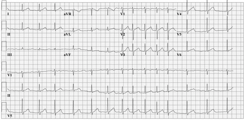 ECG
ECGDefinition
Atrial fibrillation is a rapid, irregularly irregular atrial rhythm. The atria no longer contract in an organized way; the AV node is bombarded with numerous electrical impulses and conducts them inconsistently. This produces an irregularly irregular ventricular rhythm, usually in the tachycardic range.
Classification
Types by course
- Paroxysmal: lasts less than one week and ends spontaneously or is terminated; episodes may recur
- Persistent: continuous for longer than one week
- Long-standing persistent: longer than one year, but sinus rhythm can still be restored
- Permanent: can no longer be converted to sinus rhythm, or a decision has been made not to attempt this
- Lone atrial fibrillation: without an identifiable cause in people younger than 60 years
Occurrence & epidemiology
Atrial fibrillation is one of the most common arrhythmias. Its frequency rises with age; men are affected more often than women. The lifetime risk is about 25 % for men and 20 % for women. Atrial fibrillation is responsible for about 20–25 % of all strokes.
Aetiopathogenesis
Most common causes: arterial hypertension, coronary artery disease, cardiomyopathies, valvular heart disease (mitral stenosis, mitral and tricuspid regurgitation), hyperthyroidism and binge drinking ("holiday heart"). Less common causes: pulmonary embolism, atrial septal defect and other congenital heart defects, COPD, sleep apnea, myocarditis and pericarditis.
Mechanism: early in the course, focal automaticity and micro-reentry dominate, mainly arising from muscular sleeves in the pulmonary veins. Electrical and structural remodeling of the atria then promotes functional reentrant circuits and rotors; the longer fibrillation persists, the more stable it becomes.
Because atrial contraction is absent, thrombi form, predominantly in the left atrial appendage (about 90 % of left atrial thrombi in nonvalvular atrial fibrillation). Loss of atrial contraction can reduce cardiac output at normal heart rate by as much as 20 %.
Clinical features
- Often asymptomatic; otherwise palpitations, chest discomfort, weakness, light-headedness, effort intolerance and dyspnea, particularly with a very rapid ventricular rate (often 140–160/min)
- Findings: irregularly irregular pulse, loss of a waves in the jugular venous pulse, pulse deficit (apical rate faster than the rate palpated at the wrist)
- Thromboembolism: stroke and systemic emboli to the gut, kidneys, limbs or eyes, sometimes as the first manifestation
- Heart failure, especially with a rapid ventricular rate or pre-existing reduced pump function; with sustained tachycardia, tachycardia-induced cardiomyopathy
- In pre-excitation (Wolff-Parkinson-White syndrome), atrial fibrillation may present with wide QRS complexes and a very high ventricular rate.
Diagnosis
ECG
- Absence of P waves
- Fibrillatory (f) waves between the QRS complexes, irregular in timing and morphology, at rates above 300/min; usually best seen in V1 and not apparent in all leads
- Irregularly irregular R-R intervals; usually narrow QRS complexes
- Aberrantly conducted beats after a long-short R-R sequence (Ashman phenomenon), typically with right bundle branch block morphology, can mimic ventricular extrasystoles.
Further tests
- Ambulatory ECG or event recorder when paroxysmal atrial fibrillation is suspected
- Thyroid function tests (hyperthyroidism as a cause), plus electrolytes
- Transthoracic echocardiography: structural heart disease (e.g. left atrial enlargement, wall motion abnormalities, valve disease, cardiomyopathy)
- Transesophageal echocardiography: thrombi in the atrial appendage, which are often not visible transthoracically
- Stroke risk: risk factors such as older age, hypertension, diabetes mellitus, left ventricular dysfunction and previous thromboembolism are included in scores such as the CHA₂DS₂-VA(Sc) score.
CHA₂DS₂-VA score (ESC 2024)
- Change 2024: female sex removed as Sc point.
- Point values: C 1 + H 1 + A₂ (≥ 75) 2 + D 1 + S₂ 2 + V 1 + A (65–74) 1.
Keep learning in the app
Further reading (open access)
Cross-references
More topics: Cardiology
- Acute coronary syndrome (heart attack, STEMI/NSTEMI)
- Heart failure
- Arterial hypertension (high blood pressure)
- Atrioventricular block
- Secondary hypertension
- Hypercholesterolemia and familial hypercholesterolemia
- Infective endocarditis
- Long QT syndrome and torsades de pointes
- Myocarditis
- Bundle branch block (left and right)
- Ventricular tachycardia
- Dilated cardiomyopathy
Note: Learning content for medical education – not a treatment recommendation and no substitute for diagnosis or treatment decisions in individual cases. Treatment and management are deliberately not covered on this page.