Acute coronary syndrome (heart attack, STEMI/NSTEMI)
Board exam relevance: in 26 of 105 exam reports · rank 3- Synonyms
- heart attack, myocardial infarction, MI, STEMI, NSTEMI, unstable angina, ACS
- Specialty
- Internal medicine · Cardiology
- Images
- ECG 1
- Last updated
- 10/2026 · Dr. Pascal Bafteh
Contents
Images (1)
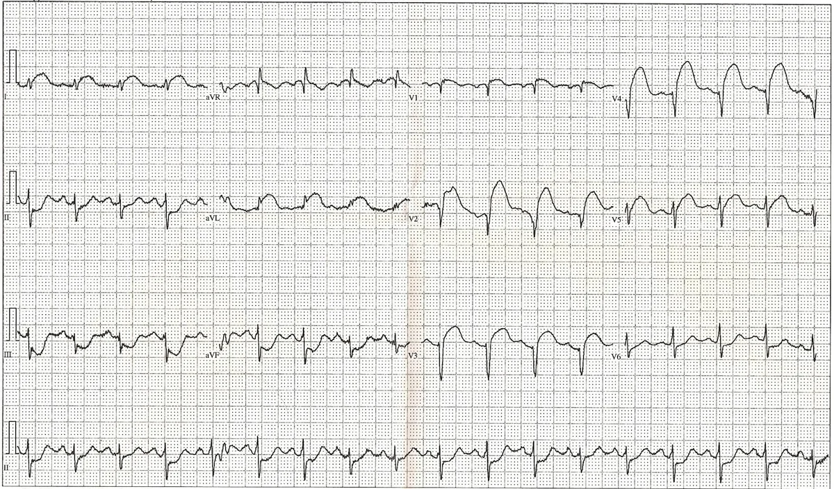 ECG
ECGDefinition
Acute coronary syndrome (ACS) comprises the acute, potentially life-threatening manifestations of coronary artery disease: ST-elevation myocardial infarction (STEMI), non-ST-elevation myocardial infarction (NSTEMI) and unstable angina.
According to the Universal Definition (most recently the Fifth Definition, 2026), myocardial infarction is present when cardiac troponin shows a rise and/or fall (at least one value above the sex-specific 99th percentile) together with evidence of acute ischemia: typical symptoms, new ischemic ECG changes or Q waves, a new regional wall motion abnormality or an intracoronary thrombus. In unstable angina troponin is not above the 99th percentile.
Classification
Types and classification
- STEMI: sustained ST elevation in at least two contiguous leads (or a STEMI equivalent) with a troponin rise; usually complete thrombotic coronary occlusion.
- NSTEMI: troponin rise without sustained ST elevation; the ECG shows ST depression, T-wave inversion or no abnormality. Together with unstable angina referred to as NSTE-ACS.
- Types of infarction: The Fifth Universal Definition (2026) replaces types 1–5 of the Fourth Definition (2018) with a clinical classification: primary infarction due to an acute coronary pathology (atherothrombosis with plaque rupture or erosion, spontaneous coronary artery dissection, coronary embolism, vasospasm); secondary infarction due to oxygen supply-demand mismatch caused by another acute condition (e.g. tachycardia, severe hypertension, hypotension, hypoxia, anemia); procedure-related infarction within 30 days of a cardiac procedure.
- MINOCA: infarction without significant coronary stenosis, e.g. due to plaque disruption without stenosis, spasm, coronary embolism or spontaneous coronary artery dissection; accounts for about 5–6 % of infarctions undergoing angiography, more often in younger women. The Fifth Universal Definition defines MINOCA as a working diagnosis ("myocardial injury with non-obstructive coronary arteries").
- Killip classification: I no signs of left ventricular failure; II mild to moderate left ventricular failure; III severe left ventricular failure with pulmonary edema; IV cardiogenic shock.
Occurrence & epidemiology
Ischemic heart disease is the leading cause of death worldwide. ACS is to be considered in men usually from about age 30 and in women usually from about age 40, earlier in diabetes mellitus. About 20 % of infarctions are silent, up to 30 % in diabetes mellitus.
Aetiopathogenesis
The underlying cause is almost always coronary atherosclerosis. Rupture of a lipid-rich, inflamed plaque with a thin fibrous cap or erosion of the plaque surface exposes thrombogenic material, followed by platelet activation and thrombus formation. Complete occlusion typically causes transmural necrosis with ST elevation, subtotal or intermittent occlusion rather subendocardial necrosis.
Risk factors: smoking, arterial hypertension, dyslipidemia (LDL cholesterol, lipoprotein(a)), diabetes mellitus, age, male sex, family history, obesity, physical inactivity and chronic kidney disease. Less common causes are spontaneous coronary artery dissection (especially younger women), coronary spasm (e.g. triggered by cocaine use), coronary embolism and vasculitides.
Clinical features
Symptoms and complications
The cardinal symptom is acute chest pain: pressing, burning or constricting, retrosternal, often radiating to the left arm, neck, jaw, back or epigastrium, persistent and not easing with rest. Dyspnea, cold sweating and nausea are common.
In inferior infarction nausea and vomiting are more frequent; second-degree AV block type Mobitz I is relatively common here, and accompanying right ventricular involvement presents with hypotension.
- Arrhythmias: ventricular tachycardia and ventricular fibrillation (especially in the first hours), atrial fibrillation, AV block
- Pump failure: acute heart failure, pulmonary edema, cardiogenic shock; in right ventricular infarction hypotension and raised jugular venous pressure with clear lungs
- Mechanical complications: ventricular septal rupture, papillary muscle rupture, free wall rupture
- Later: left ventricular aneurysm, LV thrombus with embolism, pericarditis and Dressler syndrome, ischemic cardiomyopathy
Diagnosis
ECG
- STEMI criteria (without left bundle branch block or hypertrophy): new ST elevation at the J point in two contiguous leads of at least 1 mm; in V2–V3 at least 2 mm in men aged 40 or older, 2.5 mm in men younger than 40 and 1.5 mm in women. Reciprocal ST depression supports the diagnosis.
- Evolution: hyperacute T waves, ST elevation, loss of R-wave amplitude, pathological Q waves, T-wave inversion.
- Additional leads: V7–V9 reveal a posterior infarction (circumflex occlusion), V4R right ventricular involvement.
- NSTE-ACS: ST depression, T-wave inversion or a normal ECG. Sustained ST elevation is highly specific for infarction but only moderately sensitive; serial ECGs show the typical evolution.
Localizing the infarction on the ECG
- Inferior wall: ST elevation in II, III and aVF; usually right coronary artery (ST elevation then often greater in III than in II, reciprocal ST depression in I and aVL), in left-dominant circulation also the circumflex artery
- Anterior wall (left anterior descending artery): ST elevation in the precordial leads; with proximal occlusion in V1–V6 plus I and aVL with reciprocal inferior ST depression; with occlusion of a diagonal branch more localized in V2–V3, sometimes aVL
- Lateral wall: ST elevation in I, aVL, V5 and V6 (usually circumflex artery)
- Posterior wall: horizontal ST depression in V1–V3 with tall R waves; ST elevation in the additional leads V7–V9 confirms transmural infarction (often circumflex artery)
- Right ventricle: ST elevation in the right precordial leads V3R and V4R, occasionally in V1; usually with proximal right coronary occlusion, almost always together with an inferior infarction
Laboratory tests
- High-sensitivity troponin (T or I) is the central biomarker; measured on presentation and again after 1–2 hours. A marked rise makes infarction likely; elevations without ischemia occur e.g. in heart failure, pulmonary embolism and renal failure.
Imaging and risk scores
- Echocardiography: regional wall motion abnormalities, pump function, right ventricular involvement, mechanical complications, pericardial effusion
- Coronary angiography: visualization of the infarct-related artery (occlusion, thrombus, stenosis) and the entire coronary tree
- Cardiac MRI: necrosis on late gadolinium enhancement (subendocardial to transmural), edema; important in MINOCA
- Risk scores: the TIMI score (separate versions for STEMI and NSTE-ACS) and the GRACE score are used for risk estimation; the Killip class is part of the GRACE score and of the TIMI score for STEMI, not of the TIMI score for NSTE-ACS.
Keep learning in the app
Further reading (open access)
- MSD Manual Professional: Acute Myocardial Infarction (MI)
- MSD Manual Professional: Overview of Acute Coronary Syndromes (ACS)
- StatPearls: Acute Coronary Syndrome
- StatPearls: Myocardial Infarction
- Fifth Universal Definition of Myocardial Infarction (2026), Glob Heart (PMC13523810)
- Precision Medicine for Electrocardiogram Interpretation (Rev Cardiovasc Med 2025, PMC12781007)
Cross-references
More topics: Cardiology
- Heart failure
- Atrial fibrillation
- Arterial hypertension (high blood pressure)
- Atrioventricular block
- Secondary hypertension
- Hypercholesterolemia and familial hypercholesterolemia
- Infective endocarditis
- Long QT syndrome and torsades de pointes
- Myocarditis
- Bundle branch block (left and right)
- Ventricular tachycardia
- Dilated cardiomyopathy
Note: Learning content for medical education – not a treatment recommendation and no substitute for diagnosis or treatment decisions in individual cases. Treatment and management are deliberately not covered on this page.