Aortic valve stenosis
Board exam relevance: in 1 of 105 exam reports · rank 181- Synonyms
- AS, narrowed heart valve, calcific aortic stenosis, aortic valve calcification
- Specialty
- Internal medicine · Cardiology
- Images
- Echocardiography 1 · Gross specimen 1 · X-ray 1 · ECG 1
- Last updated
- 10/2026 · Dr. Pascal Bafteh
Contents
Images (4)
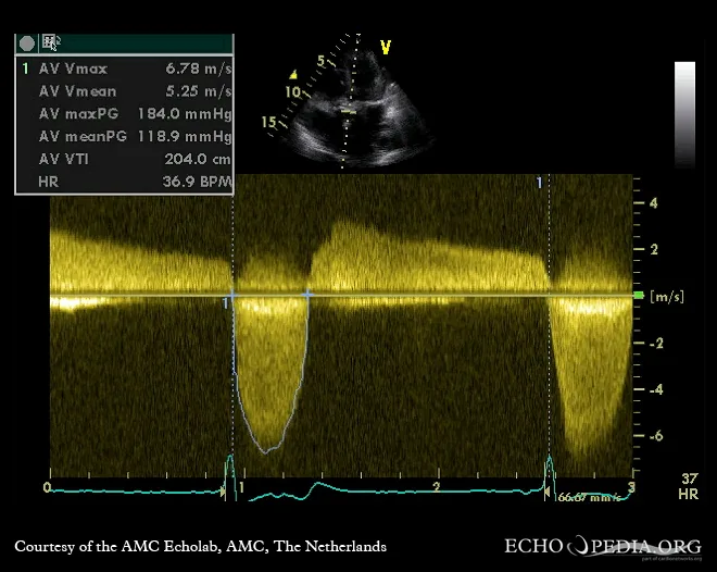 Echocardiography
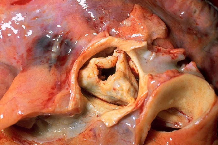 Gross specimen
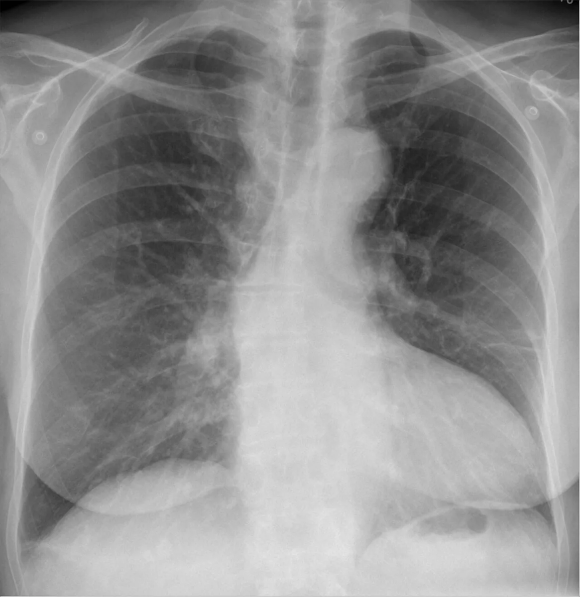 X-ray
X-ray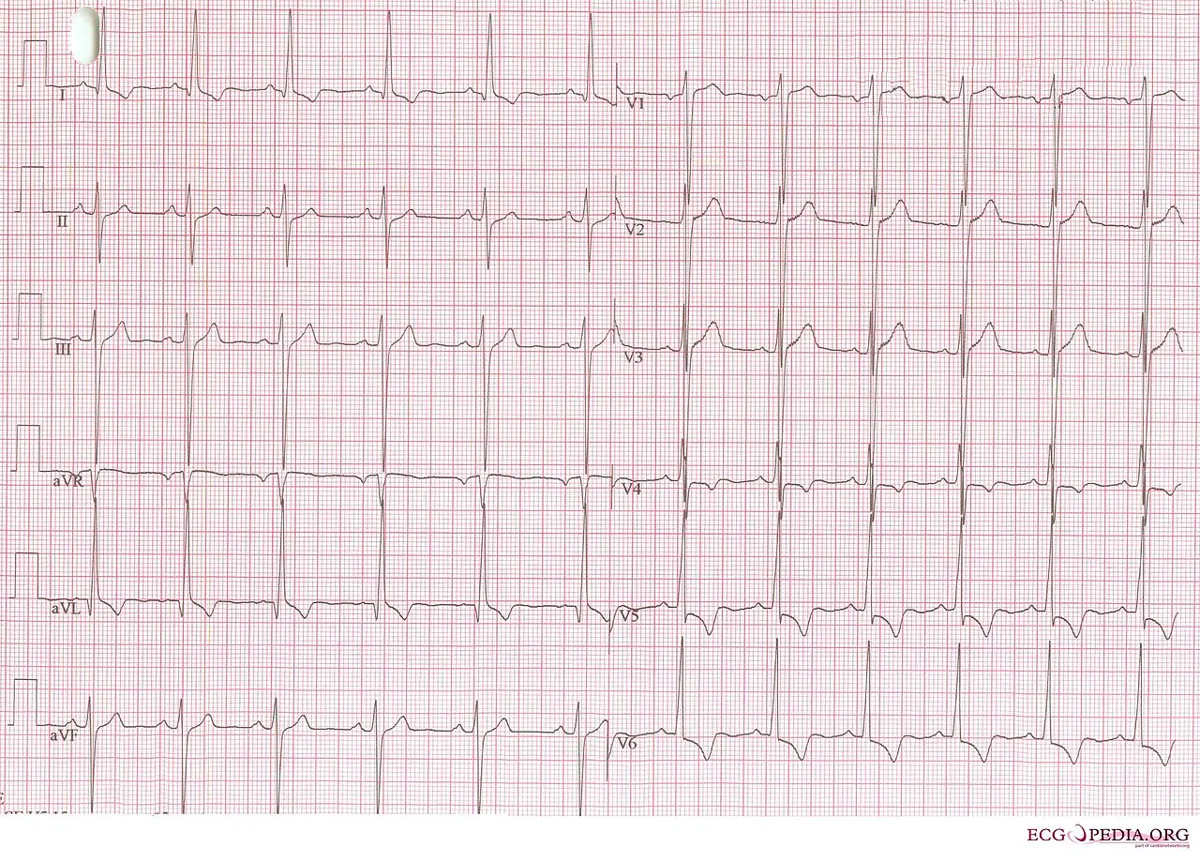 ECG
ECGDefinition
Aortic stenosis (AS) is narrowing or restricted opening of the aortic valve that obstructs blood flow from the left ventricle into the aorta during systole. The most common causes are a congenital bicuspid valve, degenerative calcification and rheumatic fever.
Aortic sclerosis describes calcium deposits on the cusps without relevant obstruction; in older people it is the most common precursor of stenosis.
Classification
Severity grades (echocardiography)
- Mild: peak velocity (Vmax) 2.5–2.9 m/s, mean gradient 10–20 mmHg or aortic valve area (AVA) 1.5–2.0 cm².
- Moderate: Vmax 3–4 m/s, mean gradient 20–40 mmHg or AVA 1.0–1.5 cm².
- Severe: Vmax of 4 m/s or more, mean gradient above 40 mmHg or AVA up to 1.0 cm².
- Very severe: Vmax above 5 m/s or mean gradient above 60 mmHg.
- Low-flow, low-gradient AS with reduced ejection fraction: AVA up to 1.0 cm², Vmax below 4 m/s, LVEF below 50 %.
- Paradoxical low-flow, low-gradient AS: AVA up to 1.0 cm², Vmax below 4 m/s, LVEF of 50 % or more with a small, hypertrophied ventricle; stroke volume index below 35 ml/m².
Occurrence & epidemiology
Aortic sclerosis is found in 25–45 % of adults over 65 years. Congenital aortic valve disease, including the bicuspid valve, affects 0.5–2 % of people, more often men. Below the age of 70 the bicuspid valve is the most common cause of AS, in older people aortic sclerosis.
Aetiopathogenesis
Calcific AS: an inflammation-mediated process similar to, but distinct from, atherosclerosis. Lipid deposition and inflammation initially cause fibrosis and calcification of the cusps without obstruction. Risk factors are arterial hypertension, smoking, hypercholesterolemia and a bicuspid valve; raised lipoprotein(a) is involved in pathogenesis and associated with faster progression.
Bicuspid aortic valve: a congenital malformation associated with coarctation of the aorta and progressive dilatation of the ascending aorta.
Pathophysiology: chronic pressure overload leads to left ventricular hypertrophy. High shear stress across the narrowed valve degrades large von Willebrand multimers; the resulting coagulopathy can cause gastrointestinal bleeding from angiodysplasia (Heyde syndrome).
Clinical features
Symptoms
AS remains asymptomatic for a long time. Typical late symptoms are:
- exertional dyspnea and heart failure
- exertional angina pectoris – due to coexisting coronary artery disease or to hypertrophy-induced ischemia even without coronary stenoses
- syncope on exertion, because cardiac output cannot be increased sufficiently
- arrhythmias up to ventricular fibrillation with sudden cardiac death
Physical findings
- pulsus parvus et tardus: small, slow-rising carotid and peripheral pulses
- sustained apical impulse due to hypertrophy; in severe AS occasionally a palpable systolic thrill
- harsh crescendo-decrescendo ejection murmur at the upper sternal border radiating to the right clavicle and both carotids; louder with leg raising and squatting, softer with the Valsalva maneuver and isometric handgrip
- second heart sound single or soft, possibly paradoxically split; a normally split second heart sound reliably excludes severe AS
- early systolic ejection click with a bicuspid valve; in older patients sometimes a musical murmur at the apex (Gallavardin phenomenon)
Diagnosis
- Echocardiography: confirms the diagnosis; shows valve morphology and cause, the extent of hypertrophy and systolic dysfunction and associated valve lesions; Doppler measurement of velocity, gradient and AVA for grading.
- Low-level pharmacological stress echocardiography: in reduced LVEF distinguishes true low-gradient AS from pseudo-stenosis.
- CT: aortic valve calcium score; severe AS is likely above 2000 Agatston units in men or above 1300 in women, and very likely above 3000 or above 1600, respectively.
- ECG: signs of left ventricular hypertrophy, with or without an ischemic ST-T pattern.
- Chest X-ray: calcification of the cusps (best on the lateral view), heart size normal or only mildly enlarged, possibly signs of heart failure.
- Exercise ECG in asymptomatic severe AS: may elicit angina, dyspnea or a fall in blood pressure.
Keep learning in the app
Further reading (open access)
Cross-references
More topics: Cardiology
- Acute coronary syndrome (heart attack, STEMI/NSTEMI)
- Heart failure
- Atrial fibrillation
- Arterial hypertension (high blood pressure)
- Atrioventricular block
- Secondary hypertension
- Hypercholesterolemia and familial hypercholesterolemia
- Infective endocarditis
- Long QT syndrome and torsades de pointes
- Myocarditis
- Bundle branch block (left and right)
- Ventricular tachycardia
Note: Learning content for medical education – not a treatment recommendation and no substitute for diagnosis or treatment decisions in individual cases. Treatment and management are deliberately not covered on this page.