Acute pericarditis
Board exam relevance: in 2 of 105 exam reports · rank 142- Synonyms
- pericarditis, inflammation of the heart sac, myopericarditis
- Specialty
- Internal medicine · Cardiology
- Images
- ECG 2
- Last updated
- 10/2026 · Dr. Pascal Bafteh
Contents
Images (2)
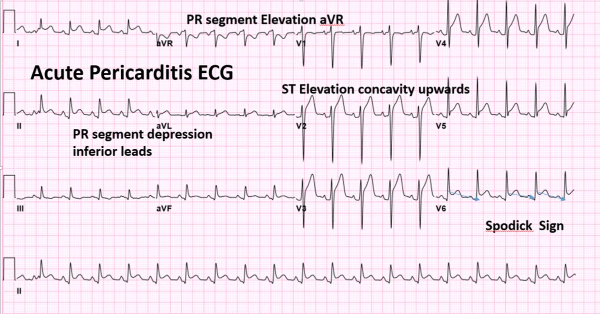 ECG
ECG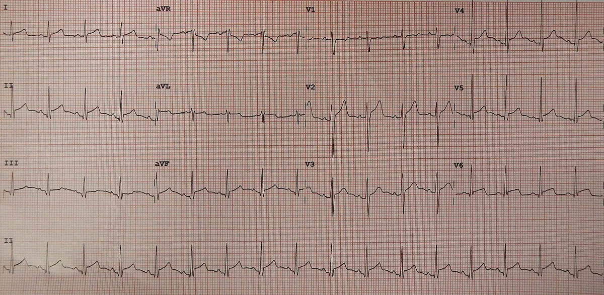 ECG
ECGDefinition
Acute pericarditis is rapidly developing inflammation of the pericardial sac, frequently with a pericardial effusion. If the inflammation extends to the adjacent myocardium, the condition is called myopericarditis.
Classification
Clinical course
- Acute: symptoms up to 4 weeks.
- Subacute: symptoms over 4 weeks to 3 months.
- Chronic: duration over 3 months.
- Incessant: symptoms persist for longer than 4–6 weeks without a symptom-free interval.
- Recurrent: new episode after a symptom-free interval of 4–6 weeks.
ECG stages
- Stage I — ST pattern: diffuse concave ST elevation + PR depression.
- Stage II: ST normalization + T flattening.
- Stage III: T inversion.
- Stage IV: normalization.
Aetiopathogenesis
- Infectious: in North America and Western Europe mostly viral or idiopathic (presumably viral), e.g. coxsackie B, echo and influenza viruses, SARS-CoV-2, HIV; in endemic regions tuberculosis is the most common cause; purulent bacterial pericarditis is uncommon, e.g. after endocarditis, pneumonia, septicemia or penetrating trauma.
- After myocardial infarction: early post-infarction pericarditis (1–2 % of infarct patients in one cohort) and the now very rare post-myocardial infarction syndrome (Dressler syndrome).
- Post-cardiac injury: post-pericardiotomy syndrome after 5–30 % of pericardiotomies; traumatic and iatrogenic pericarditis, e.g. after catheter procedures or pacemaker placement.
- Systemic diseases: rheumatoid arthritis, systemic lupus erythematosus, systemic sclerosis, inflammatory bowel disease, sarcoidosis, amyloidosis, rheumatic fever.
- Others: tumors (e.g. breast and lung cancer, leukemia), uremia, hypothyroidism, radiogenic, certain drugs, congenital pericardial anomalies.
Clinical features
- Key findings: pleuritic chest pain, fever and a pericardial friction rub, possibly dyspnea.
- Pain: dull or sharp, precordial or retrosternal, radiating to the neck and trapezius ridge (especially the left); aggravated by thoracic movement, coughing, breathing or swallowing, relieved by sitting up and leaning forward.
- Friction rub: triphasic or systolic-diastolic, often only intermittently audible.
- Accompanying features: tachypnea, non-productive cough, fever, chills, weakness.
- Course: recurrences occur in 15–30 % of idiopathic cases; rarely tamponade is the first manifestation.
Diagnosis
Diagnostic criteria
Classically the diagnosis requires at least 2 of 4 features: typical chest pain, pericardial friction rub, ECG changes, pericardial effusion. Newer criteria start from a typical clinical presentation and count additional findings:
- pericardial friction rub
- diffuse PR depression and/or ST elevation on the ECG
- raised CRP or raised ESR
- new or worsening pericardial effusion on imaging
- pericardial edema or late gadolinium enhancement on cardiac MRI
Definite with a typical presentation and at least 2 additional findings, possible with 1 additional finding, unlikely with no additional finding.
Investigations
- ECG: changes of ST segment, PR segment and T wave in most leads; unlike infarction no reciprocal ST depression (except in aVR and V1) and no pathological Q waves.
- Echocardiography: usually an effusion (often normal in purely fibrinous pericarditis); new wall motion abnormalities or abnormal strain suggest myocardial involvement.
- Cardiac MRI: detection and extent of pericardial inflammation; extension to the myocardium in up to 15–24 % of cases.
- Laboratory: leukocytosis, CRP and ESR; troponin is often raised due to epicardial inflammation, very high levels suggest myopericarditis.
- Search for the cause: chest X-ray or CT, blood count, autoimmune tests, HIV testing where appropriate; if tuberculosis is suspected, an interferon-gamma release assay, although only culture of pericardial fluid confirms or excludes the diagnosis; cytology and culture of the fluid or pericardial biopsy when the cause is unclear.
Keep learning in the app
Further reading (open access)
Cross-references
More topics: Cardiology
- Acute coronary syndrome (heart attack, STEMI/NSTEMI)
- Heart failure
- Atrial fibrillation
- Arterial hypertension (high blood pressure)
- Atrioventricular block
- Secondary hypertension
- Hypercholesterolemia and familial hypercholesterolemia
- Infective endocarditis
- Long QT syndrome and torsades de pointes
- Myocarditis
- Bundle branch block (left and right)
- Ventricular tachycardia
Note: Learning content for medical education – not a treatment recommendation and no substitute for diagnosis or treatment decisions in individual cases. Treatment and management are deliberately not covered on this page.