Brugada syndrome
Board exam relevance: in 2 of 105 exam reports · rank 142- Synonyms
- Brugada, Brugada pattern, Brugada ECG, SCN5A, channelopathy, sudden unexplained nocturnal death
- Specialty
- Internal medicine · Cardiology
- Images
- ECG 1
- Last updated
- 10/2026 · Dr. Pascal Bafteh
Contents
Images (1)
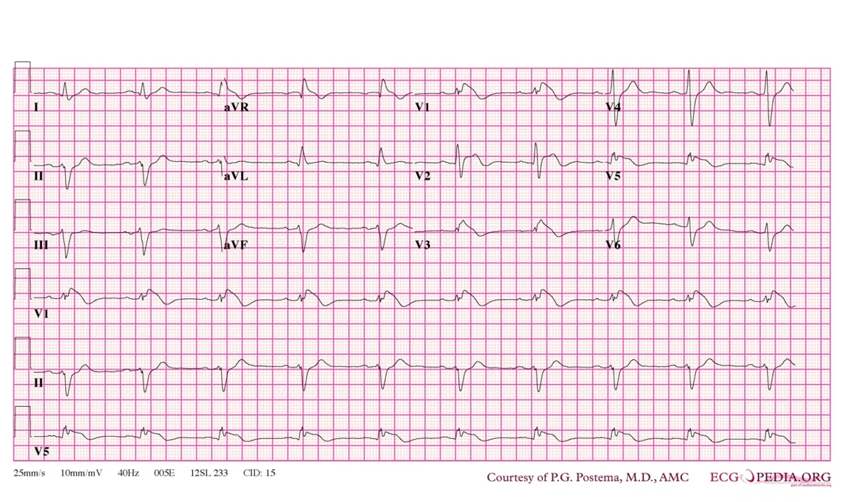 ECG
ECGDefinition
Brugada syndrome is an inherited channelopathy with characteristic ECG changes in the right precordial leads (prominent J wave, coved ST elevation and inverted T wave in V1–V3). It increases the risk of ventricular tachycardia and ventricular fibrillation with syncope and sudden cardiac death, typically without clinically evident structural heart disease.
Classification
ECG patterns
- Type 1 pattern: prominent J-point and ST elevation in V1 and V2 (sometimes V3) resembling right bundle branch block; the ST segment is coved and descends into an inverted T wave. Only a spontaneous type 1 pattern is considered diagnostic.
- Type 2 and type 3 patterns: less pronounced and not diagnostic; they may change to a type 1 pattern spontaneously, with fever or in response to drugs.
- Scores (Shanghai score) incorporating clinical, family and genetic criteria have been developed for borderline cases.
Occurrence & epidemiology
The frequency is about 5 per 10,000 people, with wide variation by ethnicity and region. 80–90 % of clinically affected people are male. In young men Brugada syndrome may account for up to about one third of sudden cardiac deaths.
Aetiopathogenesis
The cause is mutations that reduce inward sodium or calcium currents or increase early outward potassium currents. This leads to early loss of the action potential plateau, particularly in the epicardial cells of the right ventricular outflow tract. Most known mutations affect the SCN5A gene (loss of function of the sodium channel). Microscopic structural abnormalities in the epicardium of the outflow tract are often present; overlap with LQTS type 3, early repolarization syndrome and arrhythmogenic right ventricular cardiomyopathy has been described.
Triggers of arrhythmias: fever and certain drugs (sodium channel blockers, some antidepressants and antipsychotics, lithium) as well as alcohol and cocaine.
Clinical features
Most affected people are asymptomatic. In some, polymorphic VT or ventricular fibrillation causes syncope or sudden cardiac death, often at night during sleep and usually not with exercise. About 10–20 % develop atrial tachyarrhythmias, mainly atrial fibrillation, which may also be the first manifestation.
Diagnosis
- Suspicion in unexplained cardiac arrest or syncope, or a corresponding family history, without structural heart disease
- ECG: detection of the type 1 pattern; recording V1 and V2 additionally from the second and third intercostal spaces increases sensitivity
- Pharmacological provocation test with a sodium channel blocker to unmask a type 1 pattern; an ECG during fever may also reveal the pattern
- Genetic testing: yield about 20 %; evaluation of family members
Keep learning in the app
Further reading (open access)
Cross-references
More topics: Cardiology
- Acute coronary syndrome (heart attack, STEMI/NSTEMI)
- Heart failure
- Atrial fibrillation
- Arterial hypertension (high blood pressure)
- Atrioventricular block
- Secondary hypertension
- Hypercholesterolemia and familial hypercholesterolemia
- Infective endocarditis
- Long QT syndrome and torsades de pointes
- Myocarditis
- Bundle branch block (left and right)
- Ventricular tachycardia
Note: Learning content for medical education – not a treatment recommendation and no substitute for diagnosis or treatment decisions in individual cases. Treatment and management are deliberately not covered on this page.