Naevus depigmentosus (achromic naevus)
- Synonyms
- Naevus achromicus, Nävus depigmentosus, achromer Nävus, Nevus depigmentosus, achromic naevus, achromic nevus, non-pigmented naevus, hypochromic naevus
- Specialty
- Dermatology · Pigmentation disorders
- Images
- Clinical 1
- In the app
- 2 flashcards
- Last updated
- 10/2026 · Dr. Pascal Bafteh
Contents
Images (1)
Definition
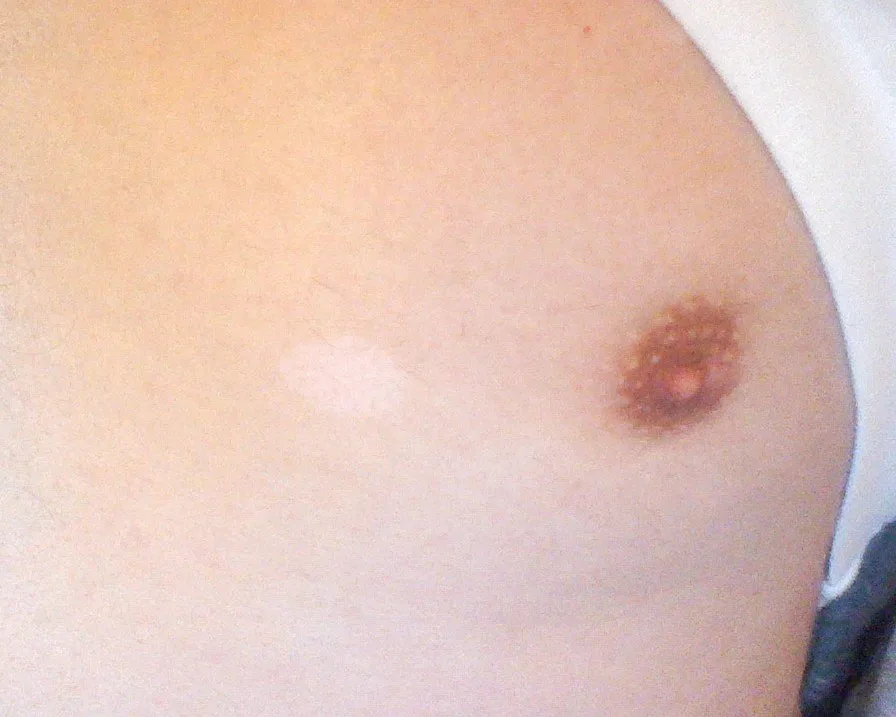
Naevus depigmentosus (achromic naevus) is a congenital or early-childhood, non-progressive hypopigmented patch that keeps its relative size and distribution throughout life and simply grows in proportion with the child. It is regarded as a form of cutaneous mosaicism. Unlike vitiligo, the patch is not completely devoid of pigment but only hypopigmented.
Classification
- Isolated type: a single circumscribed patch.
- Segmental type: sharp demarcation at the body midline with poorly defined lateral borders.
- Systematised (linear) type: follows Blaschko lines and overlaps clinically with hypomelanosis of Ito.
Occurrence & epidemiology
In a series of 67 patients the lesions were present before the age of 3 years in 92.5%; 59.7% had the isolated and 40.3% the segmental type; the back and buttocks were most commonly affected, followed by the chest and abdomen. In children with fair skin the patch may only become apparent in mid-childhood.
Clinical features
- Well-defined, off-white patch, often several centimetres across, with an irregular, sometimes serrated border; smaller satellite macules are common at the edge (like scattered splashes of paint).
- Mainly on the trunk; solitary in about half of cases.
- No change in skin texture or sensation, no dark border.
Diagnosis
- Clinical, using Coupe's criteria: present from birth or early childhood, unchanged location, no change in texture or sensation, no hyperpigmented border.
- Wood lamp: off-white, not chalk-white as in vitiligo.
- Dermoscopy: white structureless areas with a serrated border and pseudopod-like extensions, with a faint but preserved reticular pigment network.
- Biopsy (rarely needed): normal or slightly reduced melanocyte numbers with decreased melanin.
Differential diagnoses
- Vitiligo
- Hypomelanosis of Ito (incontinentia pigmenti achromians)
- Oculocutaneous albinism
- Piebaldism
- Pityriasis alba
- Nevus anemicus
- Idiopathic guttate hypomelanosis
- Progressive macular hypomelanosis
Keep learning in the app
References (selection)
Cross-references
Mentioned in
Note: This page is intended for medical education and does not replace diagnosis or treatment decisions in individual cases.