Diabetic foot ulcer (malum perforans)
- Synonyms
- Malum perforans, diabetischer Fuß, diabetisches Fußsyndrom, neuropathisches Ulkus, diabetic foot, diabetic foot ulcer, neuropathic ulcer
- Specialty
- Dermatology · Vascular skin disorders
- Images
- Clinical 6
- In the app
- 2 flashcards
- Last updated
- 10/2026 · Dr. Pascal Bafteh
Contents
Images (6)
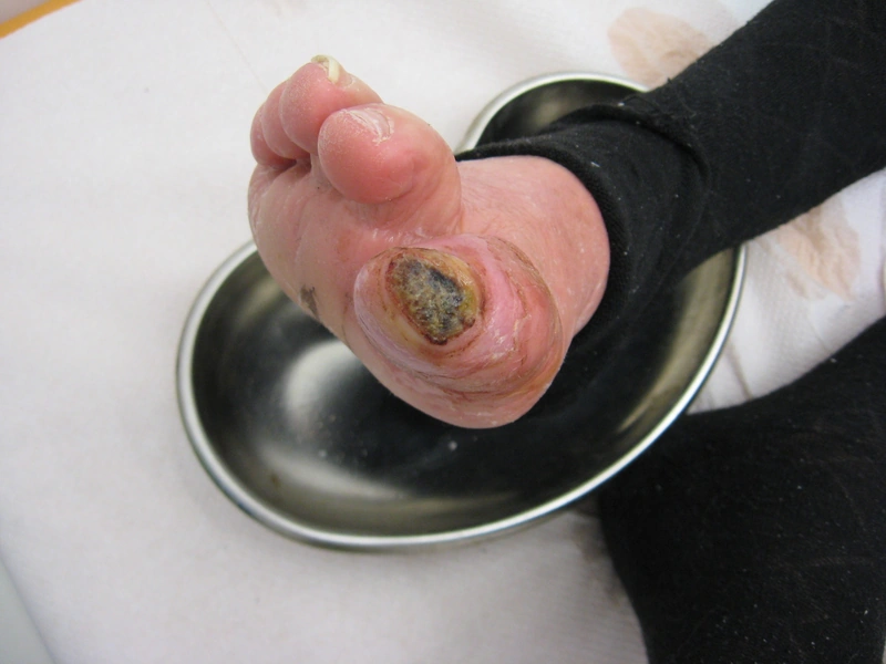
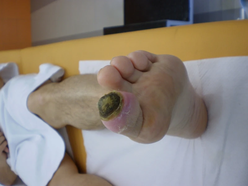
Definition
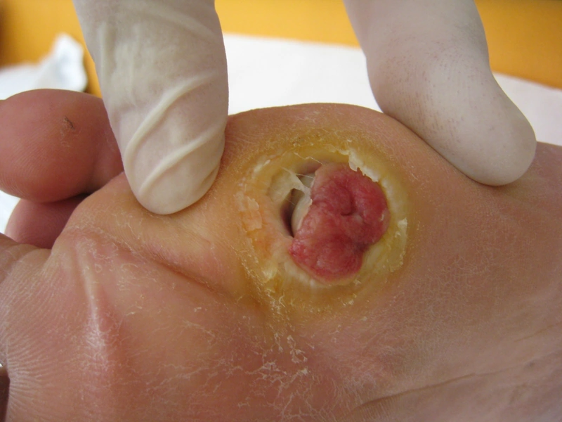
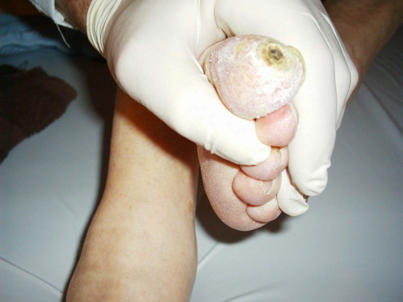
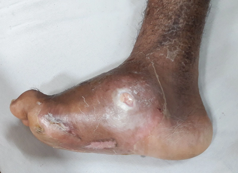
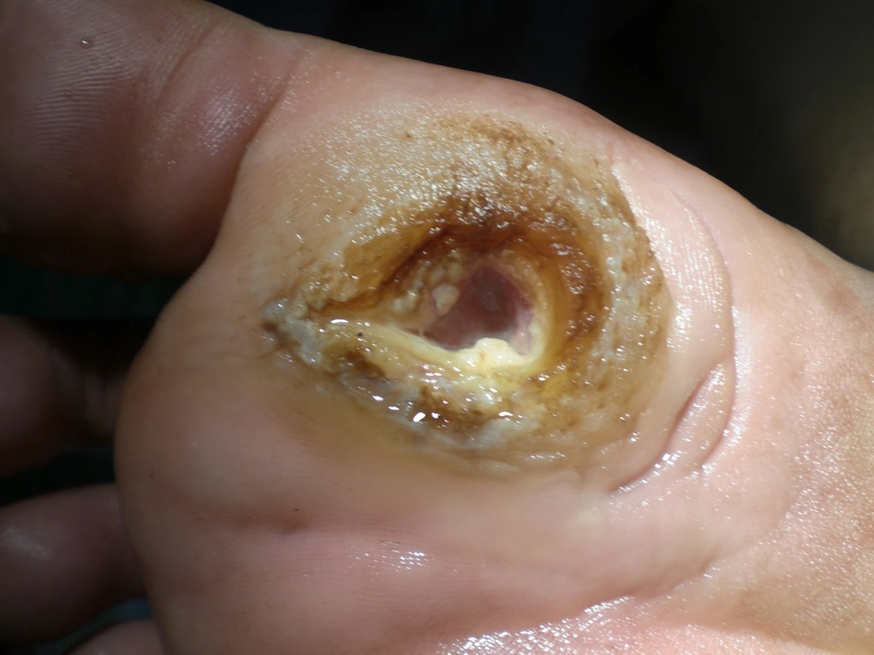
A diabetic foot ulcer is a chronic wound on the foot of people with diabetes mellitus. The main causes are diabetic polyneuropathy with loss of sensation and peripheral arterial disease. The neuropathic pressure ulcer on the sole is called malum perforans.
Occurrence & epidemiology
The annual incidence is 2–6%; up to 34% of people with diabetes develop a foot ulcer during their lifetime, and 14–24% of ulcers result in lower-extremity amputation. Risk factors are a diabetes duration of at least 10 years, poor glycaemic control (high HbA1c), male sex, previous foot ulcers, and neuropathy and circulatory disorders; ill-fitting shoes favour unnoticed injuries.
Clinical features
- Neuropathic: usually painless, circular punched-out ulcer within a callus at pressure sites (especially beneath the metatarsal heads), often preceded by a haemorrhagic blister; reduced sensation.
- Ischaemic: black necrosis up to gangrene, absent pedal pulses.
- Neuroischaemic: mixed form (about 35% in one review, neuropathic about 50%, ischaemic about 15%).
- Complications: wound infection, cellulitis, osteomyelitis; with neuropathy also Charcot arthropathy.
Diagnosis
- Classification according to the University of Texas system: grade 0–3 by depth, stage A–D by infection and ischaemia; in German-speaking countries the combined Wagner-Armstrong classification is also widely used.
- Neurological examination (monofilament, tuning fork), pulse status, ankle-brachial index (often falsely high with medial calcification, hence toe pressure), duplex ultrasound, angiography if needed.
- Probe-to-bone test and X-ray or MRI if osteomyelitis is suspected; swabs if secondary infection is suspected.
Differential diagnoses

Image credits for the thumbnails
- Venous leg ulcer: Abdullah Sarhan, “Chronisch-venöse Insuffizienz Stadium (III)”, CC BY-SA 4.0, via Wikimedia Commons · CC BY-SA 4.0 · Source
- Pyoderma gangrenosum: Monopol, “Pyoderma gangrenosum 01”, CC BY-SA 3.0, via Wikimedia Commons · CC BY-SA 3.0 · Source
- Diabetic dermopathy (shin spots): Abate, Aroucha, Nóbrega et al. (2025), “Cutaneous manifestations of diabetes mellitus: a narrative review”, CC BY 4.0, via Wikimedia Commons · CC BY 4.0 · Source
Treatment principles
- Consistent offloading, for example with offloading footwear or a total contact cast, and optimisation of glycaemic control are central.
- The wound is debrided regularly and dressed according to its healing phase; infections are treated with antibiotics, ideally after microbiological testing, and suspected osteomyelitis is investigated specifically.
- Vascular assessment is mandatory, followed by revascularisation in relevant ischaemia; care is provided by a specialised multidisciplinary foot team.
Specific treatment regimens with drugs and doses are covered in the DermaFuchs app.
Keep learning in the app
References (selection)
- DermNet: Diabetic foot ulcer
- MSD Manual Professional: Complications of diabetes mellitus
- [Diabetic foot syndrome]
- Prognostic Value of Toe Pressure Measurements in Patients with Diabetic Foot Ulcers and Medial Arterial Calcification
- Comparing the Diagnostic Accuracy of the Probe-to-Bone Test, Plain Radiography, and Serum Biomarkers in Detecting Diabetic Foot Osteomyelitis
Cross-references
Keep learning
Keep learning with flashcards, exam questions and spot diagnoses in the DermaFuchs app – free, in your browser or as an app.
Related pages: Venous leg ulcer · Pyoderma gangrenosum · Diabetic dermopathy (shin spots)
Note: This page is intended for medical education and does not replace diagnosis or treatment decisions in individual cases.