Pericardial effusion and cardiac tamponade
Board exam relevance: in 5 of 105 exam reports · rank 69- Synonyms
- fluid around the heart, pericardial tamponade, haemopericardium
- Specialty
- Internal medicine · Cardiology
- Images
- X-ray 1 · ECG 1
- Last updated
- 10/2026 · Dr. Pascal Bafteh
Contents
Images (2)
 X-ray
X-ray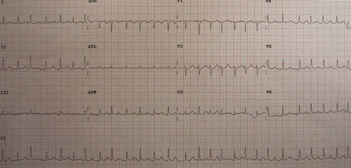 ECG
ECGDefinition
A pericardial effusion is an accumulation of fluid in the pericardial sac. Cardiac tamponade is present when a moderate or large effusion impairs filling of the heart, leading to low cardiac output and sometimes shock and death. Tamponade is primarily a clinical diagnosis.
Classification
- By composition: serous (possibly with fibrin strands), serosanguineous, blood (hemopericardium), pus, chyle.
- By distribution: free or loculated; loculated effusions and localised hematomas can cause localised tamponade of individual chambers only.
- Small effusion: under 1 cm on echocardiography, or about 100 ml.
Aetiopathogenesis
- As in pericarditis: infections, autoimmune diseases, uremia, post-infarction and post-pericardiotomy syndromes, tumors, hypothyroidism (including cholesterol pericarditis), radiogenic.
- Large chronic effusion: most often due to metastases, especially from lung and breast cancer, sarcomas, melanoma, leukemia or lymphoma.
- Hemopericardium: chest trauma, iatrogenic injury (cardiac catheterisation, pacemaker insertion, central venous line), rupture of a thoracic aortic aneurysm, rupture of the heart wall after myocardial infarction (usually 1–10 days after the infarction).
- Pathophysiology: the pericardium can stretch only slowly. If fluid (usually blood) accumulates rapidly, as little as about 150 ml can cause tamponade; with slow accumulation, up to 1500 ml are tolerated without tamponade.
Clinical features
- Pericardial effusion: often painless and asymptomatic, even when large, if it develops over months; muffled heart sounds, enlarged area of cardiac dullness; with large effusions reduced breath sounds and crackles at the left base (below the left scapula). Pulse, neck veins and blood pressure remain normal unless tamponade develops.
- Tamponade: picture similar to cardiogenic shock with low cardiac output, hypotension, tachycardia and dyspnea, but markedly distended neck veins; muffled heart sounds unless the effusion is small.
- Pulsus paradoxus: in severe tamponade almost always an inspiratory fall in systolic blood pressure of more than 10 mmHg; in advanced cases the pulse disappears on inspiration.
- Kussmaul's sign is absent in tamponade.
Diagnosis
- Echocardiography: confirms the effusion and estimates its size; signs of tamponade are exaggerated respiratory variation of transvalvular and venous flows, compression or collapse of the right heart chambers and a plethoric inferior vena cava (decrease in diameter of less than 50 % on deep inspiration).
- ECG: often reduced QRS voltage with sinus rhythm usually maintained; with large chronic effusions electrical alternans due to the "swinging heart". Low voltage and alternans are neither sensitive nor specific for tamponade.
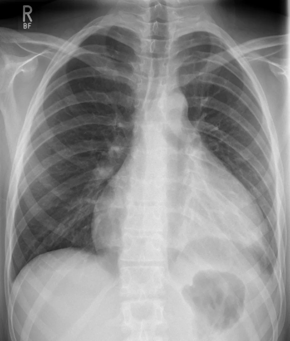
- Chest X-ray: enlarged cardiac silhouette.
- CT and cardiac MRI: effusion often an incidental CT finding (size tends to be overestimated); detection of metastases.
- Right heart catheterisation when tamponade is uncertain: diastolic pressures in all cardiac chambers and the pulmonary artery raised (about 10–30 mmHg) and equalised; in the atrial pressure curve the x descent is maintained but the y descent lost; no early diastolic dip.
- Fluid analysis: cytology and culture including acid-fast bacilli; other laboratory values of the fluid are usually non-specific.
Keep learning in the app
Further reading (open access)
Cross-references
More topics: Cardiology
- Acute coronary syndrome (heart attack, STEMI/NSTEMI)
- Heart failure
- Atrial fibrillation
- Arterial hypertension (high blood pressure)
- Atrioventricular block
- Secondary hypertension
- Hypercholesterolemia and familial hypercholesterolemia
- Infective endocarditis
- Long QT syndrome and torsades de pointes
- Myocarditis
- Bundle branch block (left and right)
- Ventricular tachycardia
Note: Learning content for medical education – not a treatment recommendation and no substitute for diagnosis or treatment decisions in individual cases. Treatment and management are deliberately not covered on this page.