Diabetic retinopathy
- Synonyms
- diabetic eye disease, NPDR, PDR, diabetic macular edema, diabetic maculopathy
- Specialty
- Internal medicine · Endocrinology & diabetes
- Images
- Clinical 2
- Last updated
- 10/2026 · Dr. Pascal Bafteh
Contents
Images (2)
Definition
Diabetic retinopathy is a microvascular complication of diabetes mellitus affecting the retina. It presents with microaneurysms, intraretinal hemorrhages, exudates, macular edema and macular ischemia and can progress to neovascularization, vitreous hemorrhage and tractional retinal detachment. It is an important cause of blindness, particularly in working-age adults.
Classification
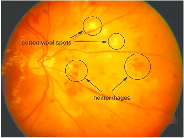
- Nonproliferative retinopathy (NPDR): develops first; increased capillary permeability, microaneurysms, dot and blot hemorrhages, hard exudates and cotton-wool spots; in later stages venous dilation and intraretinal microvascular abnormalities (IRMA).
- Proliferative retinopathy (PDR): follows NPDR and is more severe; new vessels on the optic disc or retinal surface that may grow into the vitreous.
- Diabetic macular edema: can occur with nonproliferative or proliferative retinopathy and is the most common cause of vision loss.
Occurrence & epidemiology
Over time, almost every person with diabetes develops retinopathy. Reported frequencies vary widely with the sample and method: in German studies, rates in type 2 diabetes ranged from 10.3 % to 15.7 %, and in the population-based Gutenberg Health Study (fundus photography) 21.7 %, with maculopathy in 2.3 %. In data from the North Rhine DMP (structured care program), the documented rate fell from 11.4 % (2009) to 7.6 % (2018).
Aetiopathogenesis
Chronic hyperglycemia damages the retinal capillaries through the formation of advanced glycation end products (AGE), activation of protein kinase C with endothelial dysfunction and increased permeability, and the polyol pathway with sorbitol accumulation. Capillary closure and retinal ischemia follow; the ischemic retina stimulates the growth of fragile new vessels.
Risk factors: duration of diabetes, degree of hyperglycemia and arterial hypertension. Pregnancy can worsen retinopathy.
Clinical features
For a long time there are no symptoms; even advanced retinopathy may occur without vision loss. Visual impairment results from macular edema or macular ischemia.
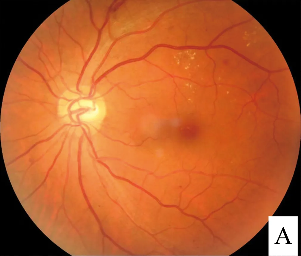
- Findings in NPDR: microaneurysms, dot and blot hemorrhages, hard exudates (yellowish, sharply defined deposits indicating chronic edema) and cotton-wool spots (white, fuzzy-edged microinfarcts of the nerve fiber layer)
- Findings in PDR: fine new vessels on the optic disc or retinal surface, preretinal fibrous tissue
- Complications: vitreous hemorrhage and tractional retinal detachment with blurred vision, floaters, flashes of light or sudden, painless vision loss; new vessels on the iris (rubeosis iridis) with neovascular glaucoma
Diagnosis
- Funduscopy with pupil dilation as the basis of diagnosis
- Color fundus photography for grading the stage of retinopathy
- Fluorescein angiography to show leakage, capillary closure and neovascularization
- Optical coherence tomography (OCT) to assess the extent and thickness of macular edema
- Slit-lamp biomicroscopy: macular edema appears as thickening and blurring of the retinal layers.
Keep learning in the app
Further reading (open access)
Cross-references
Note: Learning content for medical education – not a treatment recommendation and no substitute for diagnosis or treatment decisions in individual cases. Treatment and management are deliberately not covered on this page.