Hyperprolactinemia and prolactinoma
- Synonyms
- prolactinoma, raised prolactin, galactorrhea, pituitary adenoma, hyperprolactinemia
- Specialty
- Internal medicine · Endocrinology & diabetes
- Images
- MRI 1 · Histology 1
- Last updated
- 10/2026 · Dr. Pascal Bafteh
Contents
Images (2)
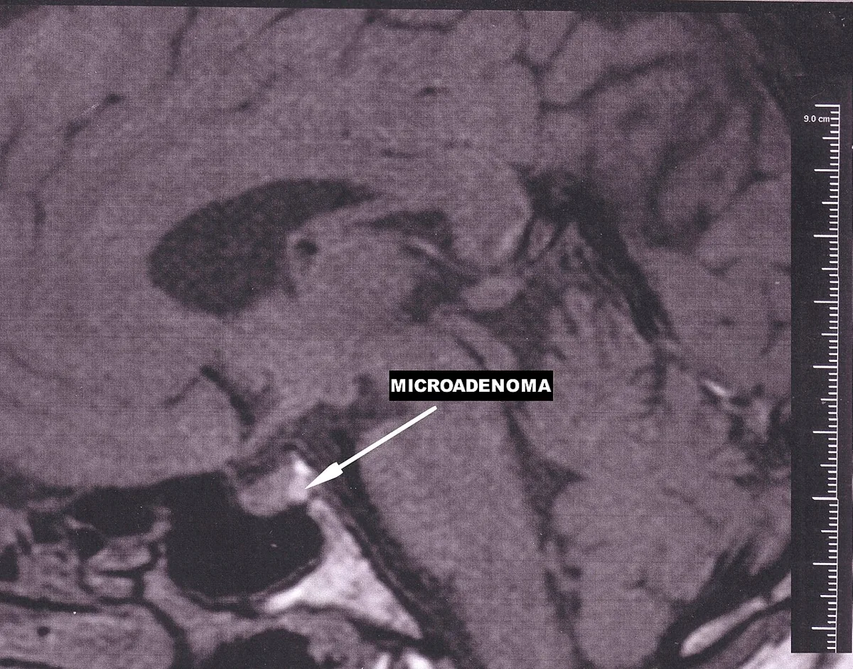 MRI
MRI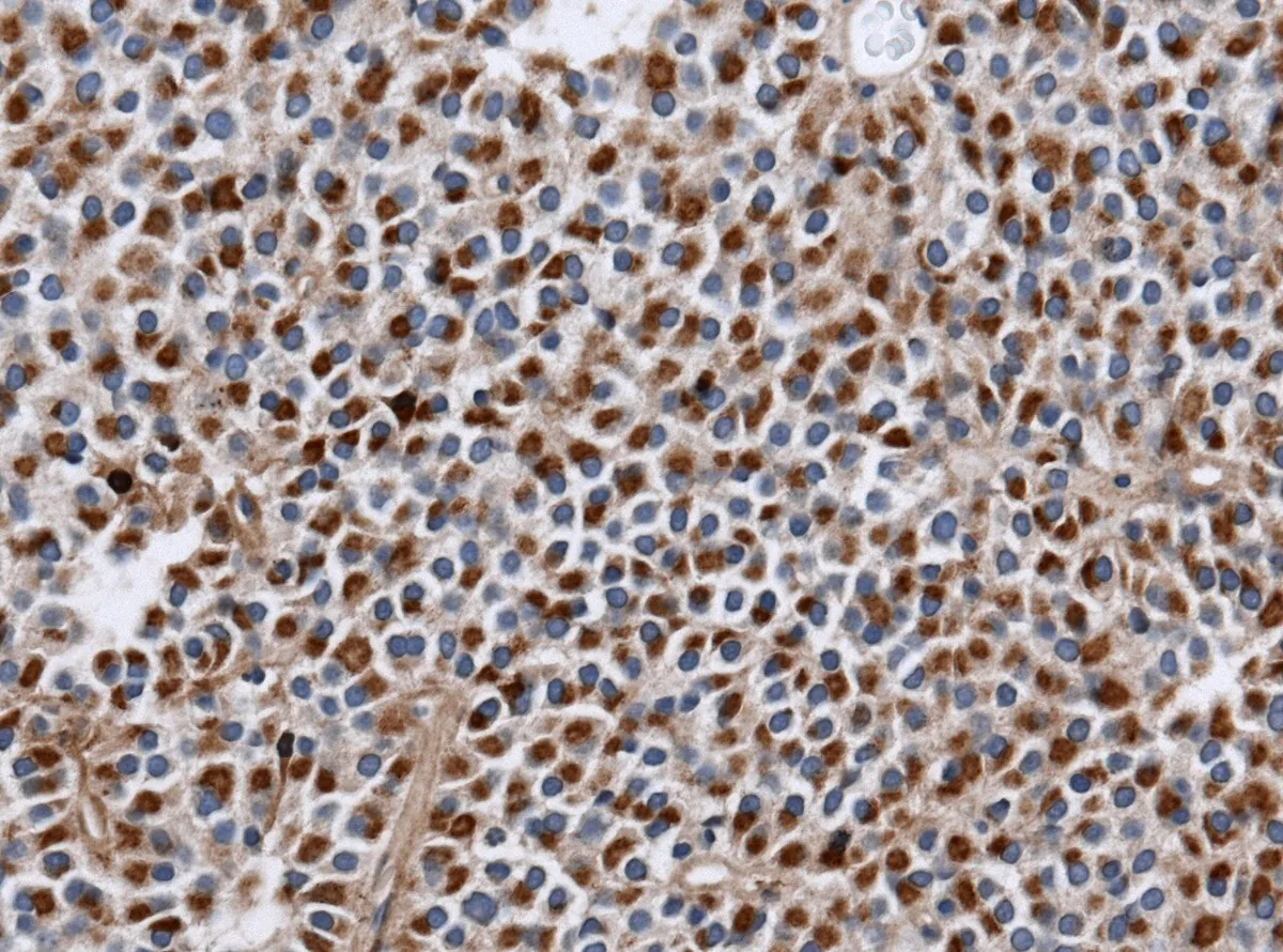 Histology
Definition
Hyperprolactinaemia is a raised blood prolactin level. Its most common pathological cause is a prolactinoma, a benign adenoma of the prolactin-producing (lactotroph) cells of the anterior pituitary. Prolactin is the hormone most frequently produced in excess by pituitary tumors. Unlike the other anterior pituitary hormones, it is regulated mainly by inhibition through hypothalamic dopamine rather than by feedback from peripheral hormones.
Classification
Size classification
- Microprolactinoma: diameter below 10 mm.
- Macroprolactinoma: diameter above 10 mm.
Occurrence & epidemiology
Lactotrophs make up about 30 % of the cells of the anterior pituitary. In adult women most prolactinomas are microadenomas at diagnosis and only a small proportion are macroadenomas. In men microadenomas are much less frequent – probably because of later recognition; prolactinomas grow more aggressively and cause mass effects more often and signs of hormone deficiency less often.
Aetiopathogenesis
Causes of hyperprolactinaemia
- Physiological: pregnancy and the postpartum period, breastfeeding or nipple stimulation, sleep, stress, food intake, hypoglycemia, early infancy.
- Pituitary: prolactinoma; compression of the pituitary stalk by other tumors (disinhibition, because dopamine no longer reaches the pituitary); empty sella syndrome; acromegaly and Cushing's disease.
- Hypothalamus: tumors, infiltration (sarcoidosis, tuberculosis, Langerhans cell histiocytosis), head injury, after encephalitis.
- Primary hypothyroidism: raised TRH increases prolactin as well as TSH secretion.
- Other organs: chronic kidney disease (reduced clearance), liver disease, chest wall lesions (scars, herpes zoster), rarely ectopic prolactin production by tumors.
- Drug-induced: including by antipsychotics and other psychoactive drugs, some blood pressure–lowering agents, opioids, H2 blockers and estrogens.
Hyperprolactinaemia inhibits the release or action of gonadotropin-releasing hormone (GnRH), leading to low gonadotropins and hypogonadism.
Clinical features
- Women: galactorrhoea (the most common sign of prolactinoma), oligomenorrhoea or amenorrhoea, anovulation and corpus luteum dysfunction with infertility; signs of estrogen deficiency such as dyspareunia and reduced libido; some women also have hirsutism.
- Men: typically headache or visual disturbances due to the mass; about two thirds have loss of libido and erectile dysfunction.
- Mass effects of macroadenomas: headache, visual field defects (typically bitemporal hemianopia due to pressure on the optic chiasm), deficiency of other anterior pituitary hormones.
Diagnosis
- Prolactin: in prolactinoma typically more than five times normal, often much higher; the level correlates with tumor size. In non-functioning masses with stalk compression it is usually not above three to four times normal.
- Exclusion of other causes: pregnancy test, medication history, kidney function; TSH and free T4 to exclude primary hypothyroidism and – with a known tumor – central hypothyroidism.
- Gonadal axis: gonadotropins and oestradiol low or normal in women, testosterone often low in men.
- Visual field testing: in all macroadenomas.
- With borderline-raised prolactin, shrinkage of the lesion under dopaminergic action can support the diagnosis.
Imaging and hook effect
- MRI sella with contrast: in confirmed hyperprolactinemia after ruling out secondary causes.
- Hook effect: at very high prolactin levels falsely low result, dilution of sample needed.
Keep learning in the app
Further reading (open access)
Cross-references
Note: Learning content for medical education – not a treatment recommendation and no substitute for diagnosis or treatment decisions in individual cases. Treatment and management are deliberately not covered on this page.