Diabetic foot syndrome
Board exam relevance: in 1 of 105 exam reports · rank 181- Synonyms
- diabetic foot, diabetic foot ulcer, DFU, malum perforans, Charcot foot, foot ulcer
- Specialty
- Internal medicine · Endocrinology & diabetes
- Images
- Clinical 3 · X-ray 1
- Last updated
- 10/2026 · Dr. Pascal Bafteh
Contents
Images (4)
 X-ray
X-rayDefinition
Diabetic foot syndrome (DFS) comprises skin changes, ulceration, infection and gangrene of the foot in people with diabetes. It is based on neuropathy and/or peripheral artery disease (PAD) together with increased susceptibility to infection, and can lead to amputation.
Classification
Wagner-Armstrong classification
Wagner grades (depth and extent of the lesion):
- Grade 0: no lesion, possibly foot deformity or cellulitis
- Grade 1: superficial ulceration
- Grade 2: deep ulcer reaching the joint capsule, tendons or bone
- Grade 3: deep ulcer with abscess formation, osteomyelitis or infection of the joint capsule
- Grade 4: limited necrosis of the forefoot or heel
- Grade 5: necrosis of the entire foot
Armstrong stages (additional findings):
- A: no infection, no ischemia
- B: infection
- C: ischemia
- D: infection and ischemia
Occurrence & epidemiology
According to the Robert Koch Institute, diabetic foot syndrome was documented in 6.2 % of people with diabetes covered by statutory health insurance in 2013 (women 5.7 %, men 6.6 %); the frequency rises almost linearly with age. In the North Rhine-Westphalia DMP (structured care program) in 2020, 9.6 % of people with type 2 diabetes had an abnormal foot status.
Aetiopathogenesis
Pathogenesis
- 3 pathogenetic pillars: neuropathy + PAD + infection.
- Neuropathic foot: warm + pink + strong pulses + painless.
- Ischemic foot: cold + livid + absent pulses.
- Charcot foot: neuropathic osteoarthropathy + 'rocker-bottom'.
- Sensory neuropathy: pressure points, foreign bodies and minor injuries are not perceived.
- Motor neuropathy: muscle atrophy leads to foot deformities such as claw and hammer toes with peak pressures under the metatarsal heads.
- Autonomic neuropathy: dry, cracked skin due to absent sweating makes it easier for pathogens to enter.
- Macroangiopathy: PAD in diabetes preferentially affects the lower-leg arteries; medial arterial calcification is common.
- Microangiopathy and impaired immunity affect wound repair and defense against infection; even minor skin defects can develop into deep, infected ulcers.
Clinical features
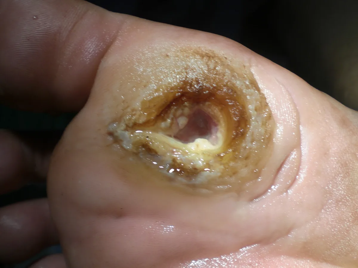
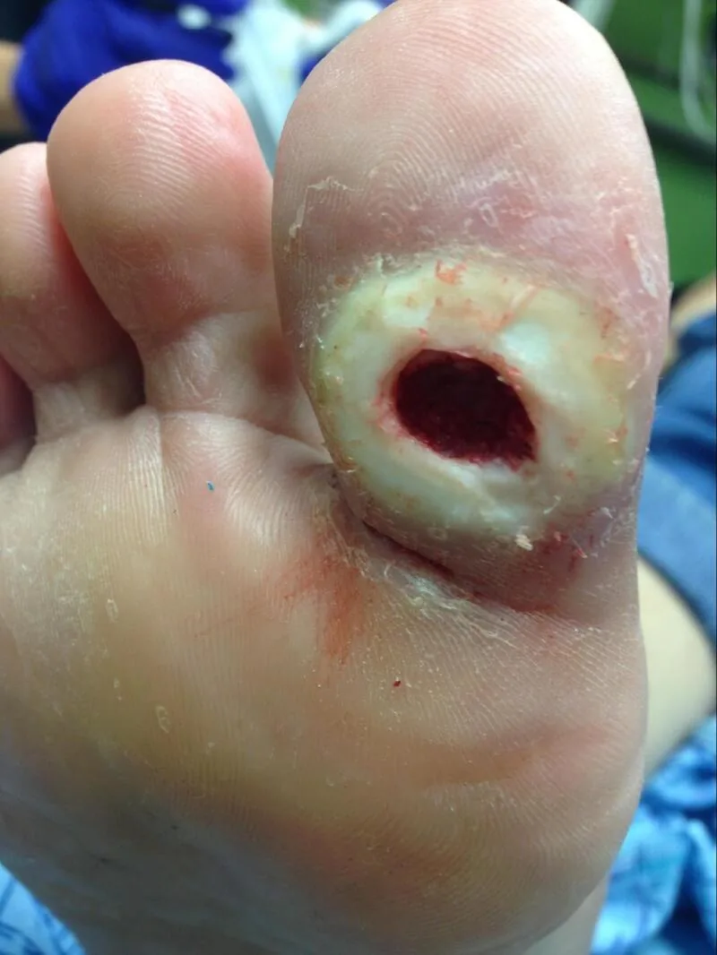
- Neuropathic ulcer (malum perforans): painless ulcer, often surrounded by callus, at pressure points, typically on the sole under the metatarsal heads; the foot is warm with palpable pulses.
- Ischemic ulcer: painful necrosis at the toes, heel or edge of the foot; the foot is cool, pale to livid, with absent pulses. With coexisting neuropathy, ischemic pain may be absent.
- Infection: redness, swelling, warmth, discharge; spread to soft tissue and bone up to osteomyelitis. Systemic signs may be absent.
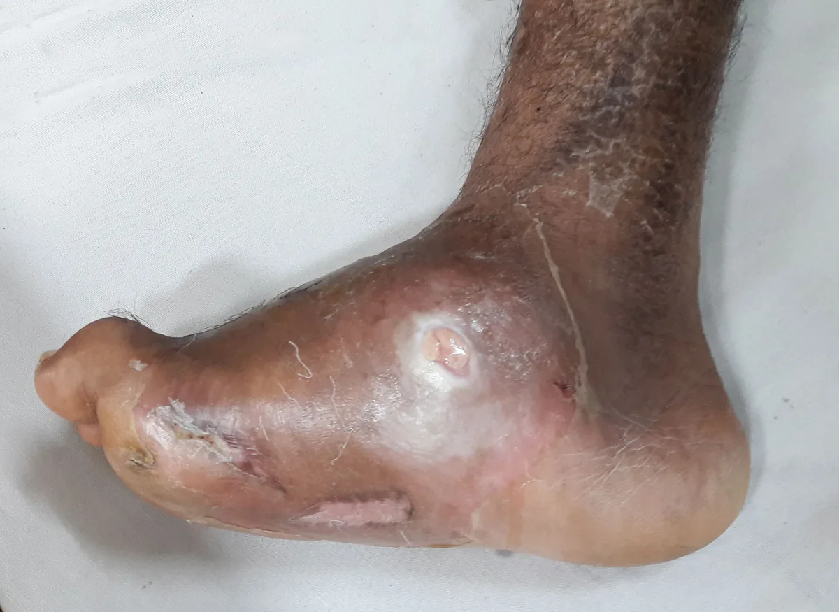
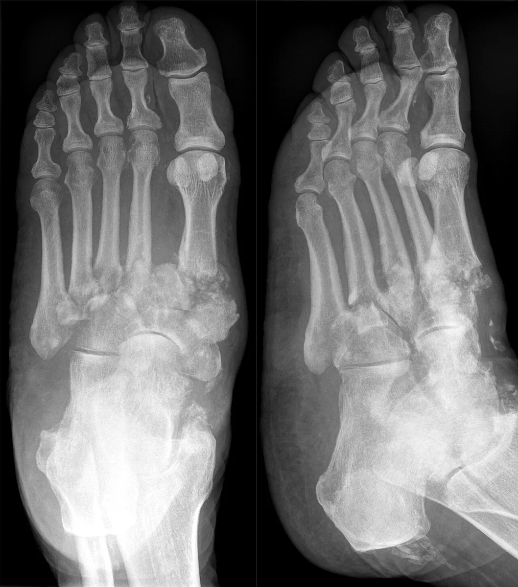
- Charcot foot: acutely warm, red, swollen and often only mildly painful foot; later fractures, subluxations and collapse of the foot arch up to rocker-bottom deformity.
- Gangrene: dry or wet-infected, localized at the toes or heel up to the entire foot.
Diagnosis
- Inspection of both feet: skin, callus, fissures, nails (onychomycosis), hair loss, deformities, ulcers with location, size and depth; footwear
- Neurological status: pressure sense with a 10 g monofilament, vibration sense, pain and temperature sensation, ankle reflexes
- Perfusion: foot pulses, ankle-brachial index (falsely high with medial arterial calcification), additionally toe pressure or toe-brachial index, transcutaneous oxygen tension and color duplex ultrasound
- Infection and bone: probing to bone (probe-to-bone test), wound swab or tissue sample to identify pathogens, foot x-ray, MRI if osteomyelitis or Charcot foot is suspected
- Laboratory: inflammatory markers, glucose metabolism and kidney function
Keep learning in the app
Further reading (open access)
Cross-references
Note: Learning content for medical education – not a treatment recommendation and no substitute for diagnosis or treatment decisions in individual cases. Treatment and management are deliberately not covered on this page.