Cutis laxa (generalised elastolysis)
- Synonyms
- Cutis laxa, generalisierte Elastolyse, Dermatochalasis generalisata, erworbene Cutis laxa, generalised elastolysis, acquired cutis laxa, lax skin
- Specialty
- Dermatology · Connective tissue diseases
- Images
- Clinical 1
- In the app
- 1 flashcards
- Last updated
- 10/2026 · Dr. Pascal Bafteh
Contents
Images (1)
Definition
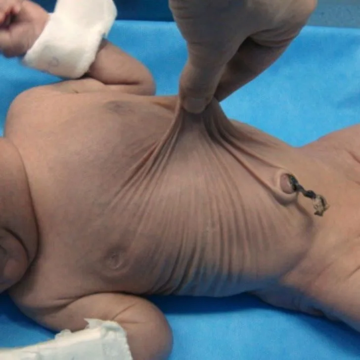
Cutis laxa is a rare disorder of elastic tissue with loose, redundant, poorly elastic skin that hangs in folds. There are inherited and acquired forms; some involve major internal organ involvement.
Classification
- Inherited: autosomal dominant, autosomal recessive and X-linked recessive inheritance; genetically heterogeneous (including mutations in elastin and fibulin genes and in ATP6V0A2, which disturbs protein glycosylation). Most inherited forms affect several organ systems.
- Acquired: not inherited, onset at any age (mean age 36.4 years in a literature review of 110 cases). Most often associated with inflammatory (43%) and neoplastic disorders (27%), and in 10% with a particular drug.
Clinical features
- Loose, inelastic, hanging skin folds with reduced recoil after stretching; the face looks prematurely aged.
- Inherited forms: most prominent around the eyes, face, neck, shoulders and thighs. Autosomal recessive forms usually start in infancy, with features such as emphysema, hernias, diverticula (oesophagus, duodenum, bladder), aortic aneurysm and skeletal abnormalities. In the autosomal dominant form the skin is often the only organ affected.
- Acquired forms: mainly in previously inflamed areas, e.g. after urticaria, angioedema or extensive inflammatory skin disease; reported associations include complement deficiency, lupus erythematosus, sarcoidosis, multiple myeloma and amyloidosis.
Histology
Dermal elastic fibres are reduced, fragmented and shortened; often hard to see on haematoxylin and eosin, but clear with an elastic stain (orcein). Giant cells engulfing elastic fibre fragments and a mild perivascular lymphocytic infiltrate may be present.
Diagnosis
- Usually a clinical diagnosis; skin biopsy with an elastic stain supports it, and electron microscopy shows fragmentation of the elastic fibres.
- If an inherited form is suspected, genetic and, where appropriate, biochemical work-up.
- In the acquired form, a targeted search for associated inflammatory and neoplastic disease.
Differential diagnoses
Keep learning in the app
References (selection)
Cross-references
Mentioned in
- Actinic reticuloid
- Ehlers-Danlos syndrome
- Limited systemic sclerosis (CREST syndrome)
- Marfan Syndrome
- Pseudoxanthoma elasticum (PXE, Grönblad-Strandberg)
- Systemic sclerosis
- Scleroedema adultorum of Buschke (scleredema)
- Ataxia-telangiectasia (Louis-Bar syndrome)
- Werner syndrome (adult progeria)
- Lipoid proteinosis (Urbach–Wiethe disease)
Note: This page is intended for medical education and does not replace diagnosis or treatment decisions in individual cases.