Acute pancreatitis
Board exam relevance: in 3 of 105 exam reports · rank 111- Synonyms
- inflammation of the pancreas, gallstone pancreatitis, biliary pancreatitis, necrotizing pancreatitis, edematous pancreatitis
- Specialty
- Internal medicine · Gastroenterology
- Images
- CT 1 · Clinical 1 · Histology 1
- Last updated
- 10/2026 · Dr. Pascal Bafteh
Contents
Images (3)
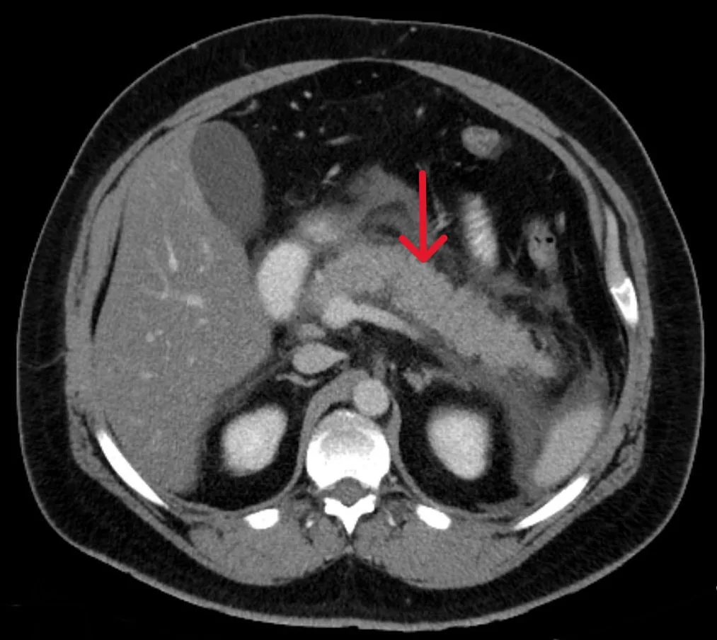 CT
CT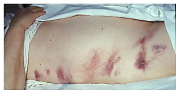
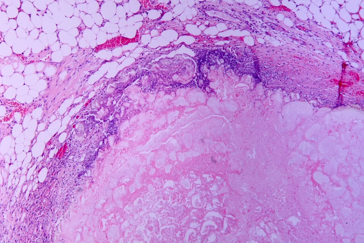 Histology
Definition
Acute pancreatitis is acute inflammation of the pancreas and sometimes of the adjacent tissues. It is caused by inappropriate release of pancreatic enzymes that injure the gland itself (autodigestion). The damaged tissue activates the complement system and the inflammatory cascade; cytokines cause inflammation and edema. In severe cases, necrosis develops with abscess, peritonitis and shock.
The diagnosis is established when at least two of the following criteria are present:
- abdominal pain consistent with the disease
- serum amylase and/or lipase more than three times the upper limit of normal
- characteristic findings on contrast-enhanced cross-sectional imaging
Classification
Atlanta classification
Local complications according to the revised Atlanta classification:
- early: acute peripancreatic fluid collection (APFC) and acute necrotic collection (ANC)
- after about 4 weeks, with capsule formation: pancreatic pseudocyst (fluid only) and walled-off necrosis (WON; fluid and necrotic material)
Types:
- interstitial edematous pancreatitis: enlarged pancreas on imaging, often with peripancreatic stranding; the most common type, mostly self-limited
- necrotizing pancreatitis: necrosis of the pancreas and/or peripancreatic tissue, best seen on contrast-enhanced cross-sectional imaging; in about 5–10% of patients, with a longer and more severe course
Severity (revised Atlanta classification 2012):
- mild: inflammation confined to the pancreas and its close vicinity, no organ failure, no local or systemic complications
- moderately severe: local or systemic complications without organ failure or with transient organ failure (resolving within 48 hours)
- severe: persistent single or multiorgan failure lasting more than 48 hours
Organ failure is assessed with the modified Marshall score (cardiovascular, renal, respiratory); a score of 2 or more in any organ system indicates organ failure.
Occurrence & epidemiology
Acute pancreatitis is common and frequently leads to a hospital stay. Most cases are due to gallstones (40–70%) and alcohol (25–35%). About 10–30% of patients develop recurrent acute pancreatitis, and about 35% of these progress to chronic pancreatitis.
Aetiopathogenesis
Gallstones: the most common cause. Pressure in the pancreatic duct probably rises because of a stone at the papilla or edema after passage of a stone; ductal hypertension leads to aberrant activation of digestive enzymes in the acinar cells. Reflux of bile acids is also discussed.
Alcohol: the second most common cause; risk generally increases with the amount, and attacks can occur in susceptible people after short periods of heavy intake. Fewer than 5% of people with chronic alcohol consumption develop acute pancreatitis. Toxic alcohol metabolites increase enzyme content, destabilize lysosomes and zymogen granules, cause calcium overload and activate stellate cells; alcohol also promotes protein plugs in the ducts.
Other causes:
- hypertriglyceridemia (about 1–4% of cases, highest risk at 1000 mg/dl or more), hypercalcemia including hyperparathyroidism
- after endoscopic imaging of the bile and pancreatic ducts (about 5–8%)
- certain drugs
- infections (e.g. coxsackievirus B, cytomegalovirus, mumps, hepatitis A and E)
- hereditary factors: an autosomal dominant mutation of the cationic trypsinogen gene causes pancreatitis in 80% of carriers; the cystic fibrosis gene also increases the risk
- structural causes: trauma, pancreatic or periampullary cancer, choledochal cyst, papillary stenosis, pancreas divisum
- methanol, smoking, pregnancy, ischemia due to hypotension or atheroembolism, tropical pancreatitis
Clinical features
- steady, boring upper abdominal pain in the epigastrium or left upper quadrant, often radiating to the back; usually sudden in gallstone pancreatitis, developing over a few days in alcoholic pancreatitis; sitting up and leaning forward relieve it, while coughing, movement and deep breathing accentuate it
- nausea and vomiting; acutely ill, sweaty appearance
- tachycardia, tachypnea and hypotension in moderate and severe cases; fever in over 60%
- limited diaphragmatic excursion, atelectasis; blunted sensorium possible
- occasionally scleral icterus due to bile duct obstruction
- ileus with decreased bowel sounds and abdominal distension; marked upper abdominal tenderness, rarely a board-like abdomen
- Grey Turner sign (flank ecchymoses) and Cullen sign (periumbilical ecchymoses) in under 3%, indicating hemorrhagic exudate
Complications: locally, fluid collections in and around the pancreas, splenic vein thrombosis, pseudoaneurysms and gastric outlet dysfunction; after about four weeks, persisting collections become encapsulated as a pseudocyst (fluid only) or walled-off necrosis (fluid and necrotic material); about one third of pseudocysts resolve spontaneously; in about one third of patients with necrosis the fluid becomes infected by gut bacteria. Systemically, shock and single or multiple organ failure (cardiovascular, respiratory, renal), favored by persistent SIRS.
Diagnosis
Laboratory tests
- Amylase and lipase rise on the first day and usually normalize after 3–5 days; lipase is more specific. Both may also be elevated in renal failure and other abdominal conditions and may remain normal when the pancreas is fibrosed after previous attacks; in hypertriglyceridemia a circulating substance can interfere with amylase measurement
- Urine trypsinogen-2 dipstick: sensitivity and specificity above 90%
- Complete blood count: white blood cells usually 12,000–20,000/µl; third-space fluid loss can raise the hematocrit to 50–55% and increase urea (BUN)
- hyperglycemia and hypocalcemia (then also check magnesium) may occur
- Liver tests: elevated bilirubin with a retained bile duct stone or compression by pancreatic edema
- anion gap metabolic acidosis in shock
- SIRS criteria (at least two): temperature above 38.3 °C or below 36.0 °C, heart rate above 90/min, respiratory rate above 20/min or PaCO2 below 32 mmHg, white blood cells above 12,000/µl, below 4,000/µl or more than 10% bands
Imaging
- Contrast-enhanced CT: the preferred imaging study to establish the diagnosis and detect local complications; early if the diagnosis is uncertain, later to detect necrosis (no contrast enhancement), fluid collections and pseudocysts
- MRI: generally preferred over CT for bile duct stones, pancreatic duct injury and necrosis; MRCP with elevated liver tests if ultrasound does not exclude duct stones
- Abdominal ultrasound if a biliary cause is suspected: gallstones, dilated bile duct; the pancreas is often obscured by bowel gas
- Abdominal X-ray: possible calcifications (indicating chronic pancreatitis), calcified gallstones, sentinel loop, colon cutoff sign
- Chest X-ray: atelectasis or pleural effusion (usually left-sided or bilateral) as signs of severe disease
Severity scores
- BISAP (1 point each, maximum 5): blood urea nitrogen (BUN) above 25 mg/dL (8.92 mmol/L), impaired consciousness (Glasgow Coma Scale below 15), SIRS, age over 60 years, pleural effusion; assessed within the first 24 hours
- CT severity index (Balthazar): based on the extent of necrosis, inflammation and fluid collections on CT; necrosis cannot yet be reliably delineated in the first 3 days after symptom onset
Keep learning in the app
Further reading (open access)
Cross-references
More topics: Gastroenterology
Note: Learning content for medical education – not a treatment recommendation and no substitute for diagnosis or treatment decisions in individual cases. Treatment and management are deliberately not covered on this page.