Type 1 diabetes mellitus
Board exam relevance: in 5 of 105 exam reports · rank 69- Synonyms
- type 1 diabetes, juvenile diabetes, insulin-dependent diabetes, T1D, LADA, autoimmune diabetes
- Specialty
- Internal medicine · Endocrinology & diabetes
- Images
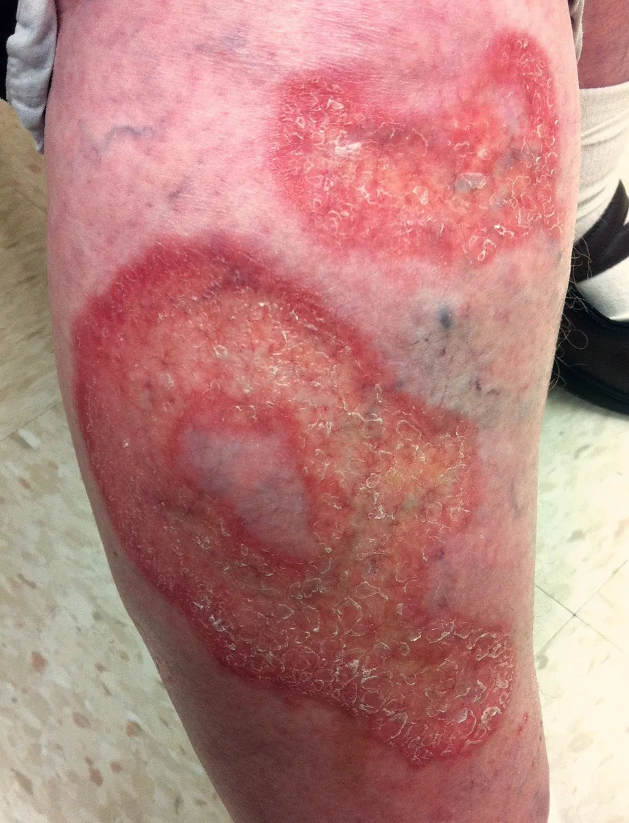
- Clinical 1 · Histology 1
- Last updated
- 10/2026 · Dr. Pascal Bafteh
Contents
Images (2)
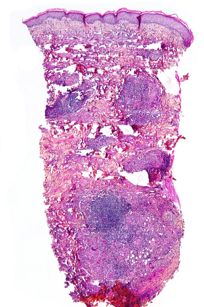 Histology
Definition
Type 1 diabetes mellitus is a chronic autoimmune disease in which the beta cells of the pancreatic islets of Langerhans are destroyed. The result is absolute insulin deficiency with chronic hyperglycemia. Type 1 accounts for about 5–10 % of all cases of diabetes.
A more slowly progressive autoimmune form in adults is called LADA (latent autoimmune diabetes in adults); because of its gradual onset it is often initially mistaken for type 2 diabetes.
Classification
Stages
- Stage 1: at least two islet autoantibodies, normal plasma glucose, no symptoms.
- Stage 2: impaired glucose tolerance or dysglycemia, still without symptoms.
- Stage 3: clinically manifest diabetes with symptoms.
Occurrence & epidemiology
Type 1 diabetes usually begins in childhood or adolescence and is the most common form of diabetes diagnosed before the age of 20. It can, however, also develop in adulthood. Its frequency differs between populations; it is particularly high, for example, in people of Scandinavian or Sardinian ancestry.
Aetiopathogenesis
In genetically susceptible individuals, an environmental factor is thought to trigger a predominantly T-cell-mediated inflammation of the islets (insulitis) that progressively destroys the beta cells. This process runs unnoticed over months to years until the remaining beta-cell mass can no longer regulate plasma glucose. The glucagon-producing alpha cells are spared.
Genetics: the most important susceptibility genes lie in the HLA complex (MHC); the haplotypes DR3-DQ2.5 and DR4-DQ8 are found in more than 90 % of those affected who are younger than 30 years. Genes outside the MHC modify the risk further.
Autoantigens: the autoantigens include glutamic acid decarboxylase (GAD), insulinoma-associated protein 2 (IA-2), zinc transporter 8 (ZnT8) and proinsulin. The islet autoantibodies detectable in serum are regarded as a consequence rather than a cause of beta-cell destruction.
Environmental factors: viral infections have been linked to disease onset (particularly coxsackieviruses and SARS-CoV-2, as well as congenital cytomegalovirus and rubella infection). Viruses may damage beta cells directly or initiate an autoimmune reaction through molecular mimicry.
Type 1 diabetes occurs more often together with other autoimmune diseases, most commonly thyroid disease, celiac disease and pernicious anemia; less commonly Addison's disease, autoimmune liver disease and myasthenia gravis.
Clinical features
When plasma glucose rises above about 160–180 mg/dl (8.9–10.0 mmol/l), glycosuria with osmotic diuresis develops. Typical features are:
- polyuria, urinary frequency and polydipsia
- weight loss, sometimes with polyphagia
- fatigue, weakness, blurred vision, nausea
- dehydration up to orthostatic hypotension; altered mental status with severe dehydration
- susceptibility to bacterial and fungal infections
In about one third of patients, diabetic ketoacidosis is the first manifestation. After onset, a prolonged phase of near-normal glucose levels may occur temporarily (honeymoon phase) because insulin secretion partially recovers.
Diagnosis
Diagnostic criteria
The criteria are valid for all forms of diabetes; with typical symptoms, one clearly elevated value is sufficient:
- HbA1c ≥ 6.5 % (≥ 48 mmol/mol)
- fasting plasma glucose ≥ 126 mg/dl (≥ 7.0 mmol/l)
- 2-hour value in the oral glucose tolerance test (75 g) ≥ 200 mg/dl (≥ 11.1 mmol/l)
- random plasma glucose ≥ 200 mg/dl (≥ 11.1 mmol/l) with symptoms of hyperglycemia
Classification as type 1
- Islet autoantibodies: antibodies against GAD, IA-2, ZnT8 and islet cells, as well as IAA, prove the autoimmune origin; in relatives they precede clinical onset.
- C-peptide: low levels indicate absent endogenous insulin secretion.
- Clinical picture: young age and ketoacidosis at onset favor type 1, but age alone does not distinguish reliably.
Keep learning in the app
Further reading (open access)
Cross-references
More topics: Endocrinology & diabetes
Note: Learning content for medical education – not a treatment recommendation and no substitute for diagnosis or treatment decisions in individual cases. Treatment and management are deliberately not covered on this page.