Primary hyperparathyroidism
Board exam relevance: in 5 of 105 exam reports · rank 69- Synonyms
- PHPT, overactive parathyroid, parathyroid adenoma, hyperparathyroidism
- Specialty
- Internal medicine · Endocrinology & diabetes
- Images
- Ultrasound 1 · Histology 1
- Last updated
- 10/2026 · Dr. Pascal Bafteh
Contents
Images (2)
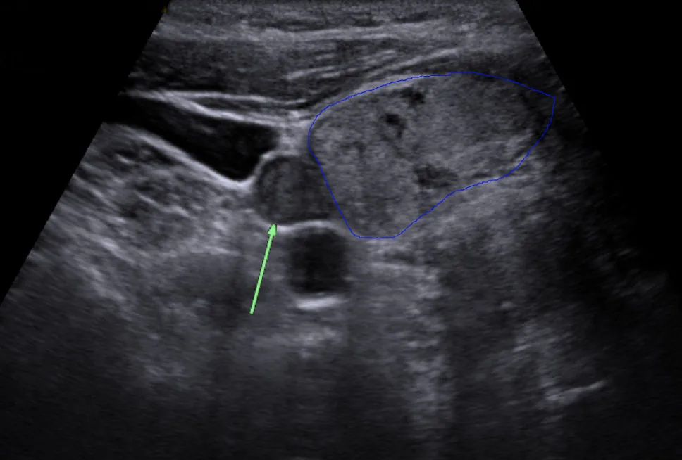 Ultrasound
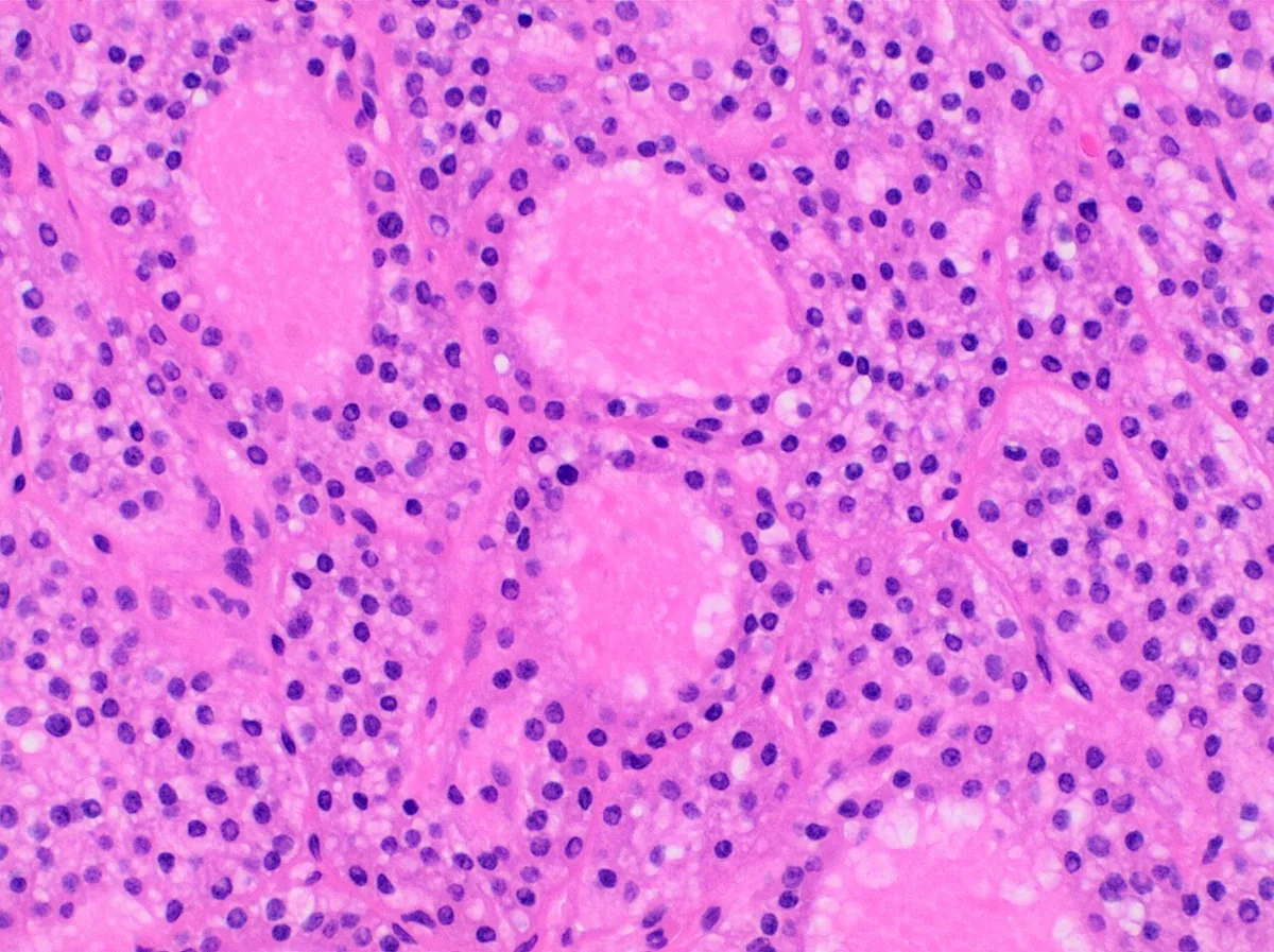 Histology
Definition
Primary hyperparathyroidism (PHPT) is excessive secretion of parathyroid hormone (PTH) by one or more parathyroid glands, largely independent of the blood calcium concentration. The consequences are hypercalcemia, hypophosphatemia and increased bone resorption. PHPT is probably the most common cause of hypercalcemia, particularly in outpatients.
Classification
Causes (histology)
- Solitary adenoma: found histologically in about 85 % of patients.
- Hyperplasia of two or more glands: about 15 %; multiglandular disease is typical of hereditary forms.
- Parathyroid carcinoma: less than 1 %; usually more severe hypercalcemia.
- Familial forms: as part of multiple endocrine neoplasia type 1 and type 2A and other hereditary syndromes.
Occurrence & epidemiology
Incidence increases with age and is higher in postmenopausal women. PHPT occurs with increased frequency three or more decades after radiation exposure of the neck. In multiple endocrine neoplasia both sexes are equally affected, onset is earlier and several glands are often involved.
Aetiopathogenesis
PTH raises serum calcium through three mechanisms:
- increased calcium reabsorption in the distal tubule
- rapid mobilisation of calcium and phosphate from bone
- increased renal formation of calcitriol and thus greater intestinal calcium absorption
Increased renal phosphate excretion causes hypophosphatemia. Persistently increased bone turnover causes osteoporosis; marked osteoclast activity produces osteitis fibrosa cystica with fibrous degeneration, cysts and "brown tumors". Long-standing hypercalcemia leads to hypercalciuria and hence kidney stones.
Clinical features
Today PHPT is usually discovered as asymptomatic hypercalcemia on routine blood tests. Symptoms are caused by hypercalcemia and its consequences:
- General and psychological: chronic fatigue, poor concentration, confusion, depression
- Gastrointestinal: anorexia, nausea, vomiting, constipation, abdominal pain
- Kidney: polyuria, nocturia and polydipsia due to impaired urinary concentration; kidney stones, often with colic
- Bone and muscle: osteoporosis, bone pain and fractures in osteitis fibrosa cystica; proximal muscle weakness and atrophy
- Heart: in severe hypercalcemia a shortened QTc interval and arrhythmias
In PHPT total calcium is rarely above 12 mg/dl (3 mmol/l). Levels of 14–15 mg/dl (3.5–3.7 mmol/l) raise concern for parathyroid carcinoma.
Diagnosis
Laboratory tests
- Calcium: repeatedly raised serum calcium; ionised calcium is almost always elevated. When protein levels are abnormal, ionised calcium is more informative.
- Intact PTH: elevated or inappropriately high-normal for the degree of hypercalcemia. In PTH-independent hypercalcemia PTH is usually below 20 pg/ml.
- Phosphate: low, especially with increased renal phosphate excretion.
- Alkaline phosphatase: often raised when bone turnover is increased.
- Urinary calcium: normal or high.
- Distinction from familial hypocalciuric hypercalcemia (FHH): low fractional calcium excretion (below 1 % in FHH, 1–4 % in PHPT), frequent hypermagnesemia, early onset, absence of symptoms and hypercalcemia without hypercalciuria in relatives; usually caused by inactivating mutations of the CASR gene.
- In chronic kidney disease: high calcium with normal phosphate suggests primary, high phosphate with low calcium suggests secondary hyperparathyroidism.
Imaging and further tests
- Bone density (DXA): additionally at the distal radius, the most common site of bone loss in hyperparathyroidism.
- Radiography: in advanced disease bone cysts, a heterogeneous skull and subperiosteal resorption at the phalanges and distal clavicle.
- Renal ultrasound: detection of kidney stones or nephrocalcinosis.
- Parathyroid localisation: high-resolution ultrasound, technetium-99m sestamibi scintigraphy (also as SPECT), contrast-enhanced 4D CT or MRI. These localise an abnormal gland but do not establish the diagnosis.
- Clues to hereditary forms: young age, multiglandular disease or other endocrine tumors; hyperparathyroidism before the age of 30 suggests MEN 1.
Keep learning in the app
Further reading (open access)
Cross-references
Note: Learning content for medical education – not a treatment recommendation and no substitute for diagnosis or treatment decisions in individual cases. Treatment and management are deliberately not covered on this page.