Pheochromocytoma
Board exam relevance: in 5 of 105 exam reports · rank 69- Synonyms
- paraganglioma, adrenal medullary tumour, catecholamine-secreting tumor, pheo
- Specialty
- Internal medicine · Endocrinology & diabetes
- Images
- Gross specimen 1 · Histology 1
- Last updated
- 10/2026 · Dr. Pascal Bafteh
Contents
Images (2)
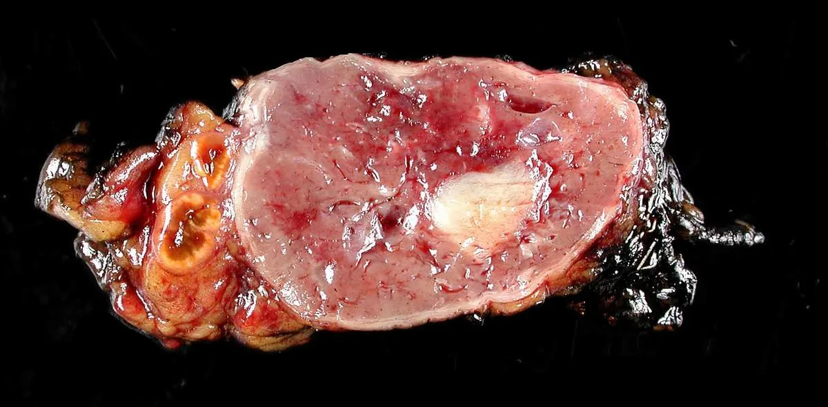 Gross specimen
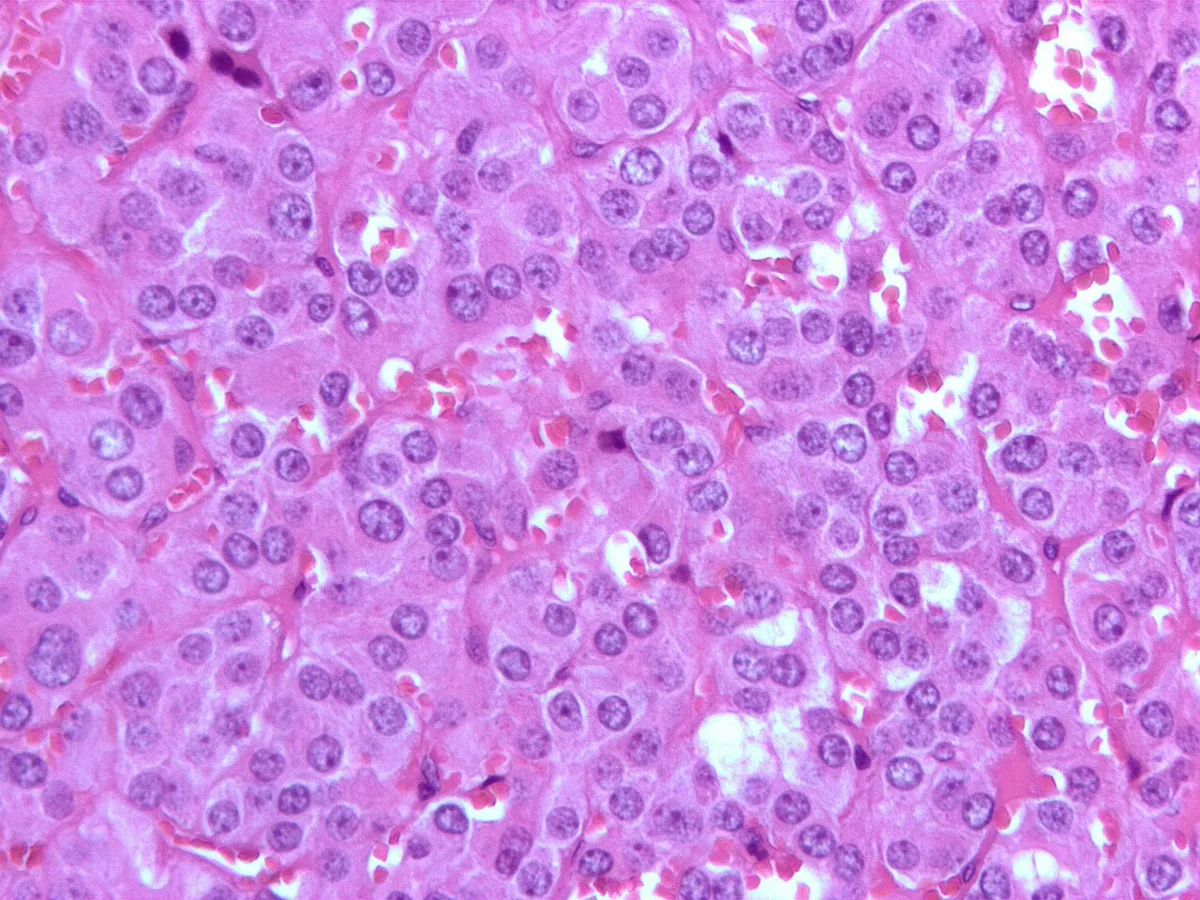 Histology
Definition
A phaeochromocytoma is a catecholamine-secreting tumor of chromaffin cells, usually in the adrenal medulla. It causes persistent or paroxysmal hypertension. Similar tumors outside the adrenal gland in the sympathetic nervous system are called paragangliomas. Head-and-neck paragangliomas arise from parasympathetic ganglia and only rarely secrete catecholamines.
Classification
- Location: about 90 % in the adrenal medulla; extra-adrenal e.g. in the paraganglia of the sympathetic chain, retroperitoneally along the aorta, in the organ of Zuckerkandl at the aortic bifurcation, in the bladder, the carotid body or the pericardium.
- Laterality: bilateral in about 10 % (20 % in children).
- Malignancy: adrenal phaeochromocytomas are malignant in about 10 %, extra-adrenal paragangliomas in about 30 %. Regardless of histology, a tumor is considered benign if it has not invaded the capsule and no metastases are present; larger tumors are more likely to recur or metastasise.
- Sporadic or hereditary: a substantial proportion is hereditary – about one third to nearly half, depending on the source.
Occurrence & epidemiology
About 0.2–0.6 % of people with hypertension have a phaeochromocytoma. Adrenal phaeochromocytomas affect both sexes equally. They occur at any age, with a peak between 20 and 40 years. The average diameter is 5–6 cm and the weight 50–200 g.
Aetiopathogenesis
The tumor releases noradrenaline, adrenaline, dopamine and dopa in varying proportions. Catecholamines cause vasoconstriction, tachycardia and metabolic effects: hyperglycemia up to diabetes mellitus through inhibition of insulin release, raised free fatty acids and a contracted plasma volume. Phaeochromocytomas in MEN 2, unlike sporadic tumors, produce disproportionately more adrenaline. A raised 3-methoxytyramine (a dopamine metabolite) indicates a more aggressive tumor.
Genetic syndromes at a glance
- Common genes: VHL + RET + NF1 + SDHB/C/D/AF2 + TMEM127 + MAX.
- MEN-2A: MTC + pheo + primary hyperparathyroidism.
- MEN-2B: MTC + pheo + marfanoid + mucosal neuromas (no hyperparathyroidism).
- VHL: pheo + hemangioblastoma + renal cell carcinoma + pancreatic cysts.
- NF1: café-au-lait + neurofibromas + Lisch nodules + pheo in ~1 %.
Clinical features
Hypertension is present in most patients and is paroxysmal in about 45 %. Common symptoms and signs:
- severe headache, sweating, palpitations and tachycardia
- cold, clammy skin, pallor
- postural hypotension
- angina, dyspnea, tachypnea
- nausea, vomiting, epigastric pain, constipation
- visual disturbances, paraesthesia, a sense of impending doom
Triggers of attacks: palpation of the tumor, postural change, abdominal pressure, induction of anesthesia, emotional stress and – with bladder tumors – micturition. In older patients, severe weight loss with persistent hypertension suggests phaeochromocytoma.
Between attacks, examination is usually normal apart from hypertension. Retinopathy and cardiomegaly are often less severe than expected for the degree of hypertension; however, a catecholamine cardiomyopathy can develop, and cardiac and cerebrovascular events are more frequent than in others with similar blood pressure.
Diagnosis
Biochemical tests
- Plasma free metanephrines: sensitivity up to 99 %; normetanephrine is elevated more often than metanephrine. Metanephrines are produced continuously, whereas adrenaline and noradrenaline are released intermittently.
- 24-hour urinary fractionated metanephrines: sensitivity about 95 %, slightly less specific than plasma. Two or three normal results while blood pressure is raised make phaeochromocytoma extremely unlikely.
- Less suitable: vanillylmandelic acid and free urinary catecholamines.
- Falsely high values: with extreme stress, sleep apnea, coma, dehydration, with certain psychoactive and illicit drugs and – for vanillylmandelic acid – after certain foods.
- Clonidine suppression test: possible with raised plasma catecholamines but rarely necessary.
- Associated findings: hyperglycemia or glycosuria, falsely high hemoglobin and hematocrit due to hemoconcentration.
Localisation and genetics
- CT or MRI of the chest and abdomen for localisation after a positive biochemical result.
- Nuclear medicine: gallium-68 DOTATATE PET is the most sensitive and is replacing MIBG scintigraphy; FDG-PET especially with succinate dehydrogenase mutations.
- Fine-needle biopsy of an adrenal mass: can precipitate an acute hypertensive crisis in phaeochromocytoma.
- Search for syndromes: café-au-lait spots (neurofibromatosis), mucosal neuromas (MEN 2B); serum calcitonin as a clue to MEN 2.
- Genetics: panel testing in all patients; the genotype determines the clinical and biochemical profile.
Keep learning in the app
Further reading (open access)
Cross-references
More topics: Endocrinology & diabetes
Note: Learning content for medical education – not a treatment recommendation and no substitute for diagnosis or treatment decisions in individual cases. Treatment and management are deliberately not covered on this page.