Thyroid cancer
Board exam relevance: in 1 of 105 exam reports · rank 181- Synonyms
- thyroid carcinoma, papillary thyroid cancer, follicular thyroid cancer, medullary thyroid cancer, anaplastic thyroid cancer
- Specialty
- Internal medicine · Endocrinology & diabetes
- Images
- Ultrasound 1 · Blood smear & cytology 1 · Histology 1
- Last updated
- 10/2026 · Dr. Pascal Bafteh
Contents
Images (3)
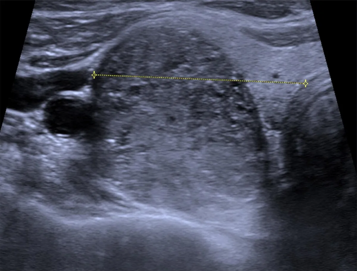 Ultrasound
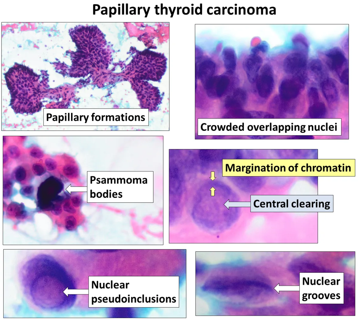 Blood smear & cytology
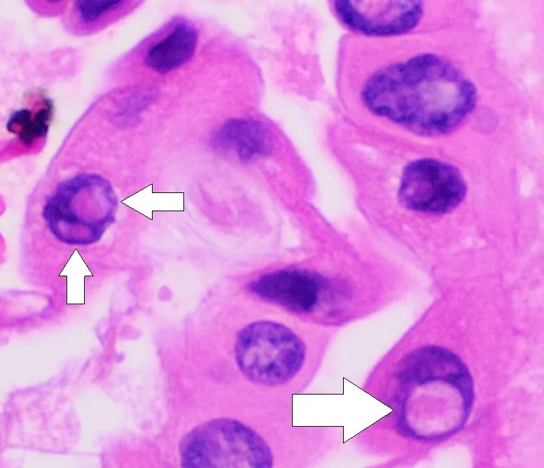 Histology
Definition
Thyroid cancers are malignant tumors of the thyroid gland. They arise either from the follicular epithelium (papillary, follicular, oncocytic, poorly differentiated and anaplastic carcinoma) or from the parafollicular C cells (medullary carcinoma).
Papillary and follicular carcinomas are grouped as differentiated thyroid cancer: they resemble normal thyroid tissue histologically and still secrete thyroglobulin. Poorly differentiated and anaplastic carcinomas are aggressive and secrete less thyroglobulin. Noninvasive follicular thyroid neoplasm with papillary-like nuclear features (NIFTP) is now considered a benign lesion.
Classification
Histological types
- Papillary carcinoma: 80–90 % of all thyroid cancers; spreads mainly via lymphatics.
- Follicular carcinoma: less common; more frequent in older people and in iodine-deficient regions; hematogenous spread with a higher risk of distant metastases.
- Oncocytic carcinoma (formerly Hürthle cell carcinoma): a distinct entity with mitochondria-rich cytoplasm; encapsulated or widely invasive.
- Anaplastic carcinoma: undifferentiated, about 1 % of thyroid cancers.
- Medullary carcinoma: about 4 %, arising from the calcitonin-producing C cells; sporadic or hereditary.
Occurrence & epidemiology
In Germany about 4,200 women and 1,800 men were diagnosed with thyroid cancer in 2022. The median age at diagnosis was 51 years in women and 55 years in men, which is relatively young for a cancer. The rise in incidence since 1999 is almost entirely due to papillary carcinoma; a plateau has now been reached. Most carcinomas are detected at an early stage (UICC I).
In papillary carcinoma the female-to-male ratio is 3:1; most patients are between 30 and 60 years old, and it is familial in 3–9 %. Anaplastic carcinoma mainly affects older people. The increase in papillary carcinoma is mostly due to small tumors found incidentally on neck imaging.
Aetiopathogenesis
- Ionising radiation: an established risk factor; the child's thyroid is particularly sensitive (e.g. uptake of radioactive iodine after reactor accidents, thyroid exposed to ionising radiation in medical settings). Tumors after radiation exposure appear from about 10 years after exposure, and the risk remains increased for 30–40 years; most of them are benign. Carcinomas after radiation exposure are often multicentric, and gene fusions are typical.
- Iodine deficiency and benign thyroid disease: goiter and adenomas are often found in the history.
- Molecular changes: BRAF V600E mutation in papillary carcinoma, RAS mutations in follicular carcinoma; extensive loss of heterozygosity and mitochondrial DNA mutations in oncocytic carcinoma.
- Hereditary medullary carcinoma: germline mutation of the RET proto-oncogene (autosomal dominant), either as familial medullary thyroid carcinoma or as part of multiple endocrine neoplasia types 2A and 2B.
Clinical features
Most thyroid cancers present as an asymptomatic nodule found incidentally on examination or imaging. Lymph node, lung or bone metastases as the presenting sign are rare. Suspicious features are a stony-hard, fixed nodule, cervical lymphadenopathy and hoarseness due to recurrent laryngeal nerve palsy.
- Papillary carcinoma: lymphatic metastases to regional lymph nodes in about one third, later also lung metastases.
- Follicular and widely invasive oncocytic carcinoma: hematogenous distant metastases, including outside the neck.
- Anaplastic carcinoma: rapid, painful enlargement of the thyroid.
- Medullary carcinoma: usually an asymptomatic nodule; metastases to cervical and mediastinal lymph nodes, occasionally to liver, lungs and bone. Ectopic production of other hormones and peptides (e.g. ACTH, vasoactive intestinal peptide, serotonin) can cause a dramatic hormonal picture. Serum calcium remains normal despite high calcitonin levels.
Histology
- Papillary carcinoma: fine, stippled psammomatous microcalcifications.
- Oncocytic carcinoma: tumor cells with mitochondria-rich, "oncocytic" cytoplasm.
- Medullary carcinoma: characteristic amyloid deposits that stain with Congo red.
- Ancillary tests on aspirate or tissue: immunostaining for thyroglobulin or BRAF V600E protein and molecular testing.
Diagnosis
- Ultrasound: features suggesting malignancy are hypoechogenicity, marked internal vascularity, irregular margins, a nodule taller than it is wide, and microcalcifications (papillary) or dense, homogeneous calcification (medullary).
- Fine-needle aspiration with cytology: the most common route to diagnosis, complemented by immunostaining and molecular tests.
- Serum calcitonin: the best laboratory test for medullary carcinoma, in which it is greatly elevated.
- Radiography or ultrasound in medullary carcinoma: dense, homogeneous, conglomerate calcification within the primary tumor.
- Genetic testing: germline testing for RET mutations in all patients with medullary carcinoma; genetic testing and calcitonin measurement in relatives. In hereditary forms a RET germline mutation is found in 98 %; the affected codons correlate with the phenotype.
- Exclusion of phaeochromocytoma in MEN 2 by measuring plasma metanephrines.
Keep learning in the app
Further reading (open access)
Cross-references
Note: Learning content for medical education – not a treatment recommendation and no substitute for diagnosis or treatment decisions in individual cases. Treatment and management are deliberately not covered on this page.