Subacute thyroiditis (de Quervain)
Board exam relevance: in 1 of 105 exam reports · rank 181- Synonyms
- de Quervain thyroiditis, subacute granulomatous thyroiditis, giant cell thyroiditis, painful thyroiditis, thyroid inflammation
- Specialty
- Internal medicine · Endocrinology & diabetes
- Images
- Ultrasound 1 · Histology 1
- Last updated
- 10/2026 · Dr. Pascal Bafteh
Contents
Images (2)
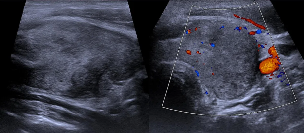 Ultrasound
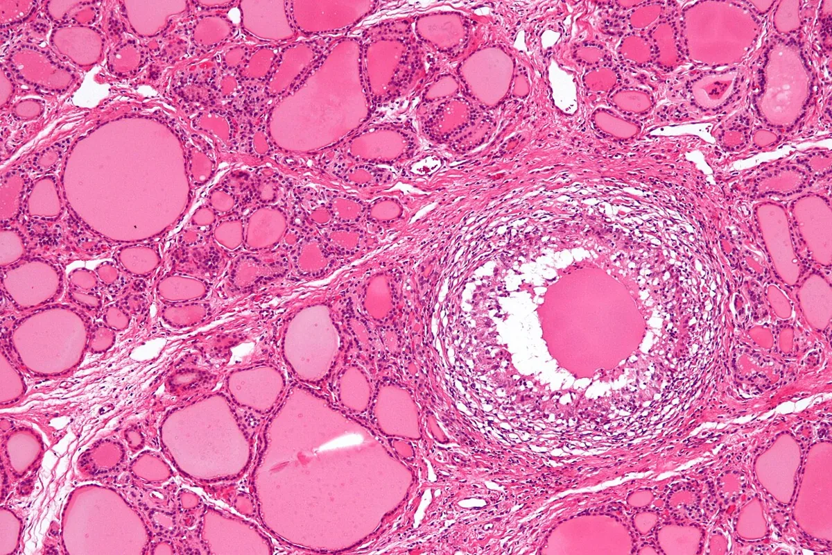 Histology
Definition
Subacute thyroiditis (de Quervain thyroiditis), also called subacute granulomatous or giant cell thyroiditis, is an acute, painful inflammation of the thyroid that is probably triggered by a viral infection. Typical features are fever, a tender neck and initial hyperthyroidism due to release of stored thyroid hormone, which may be followed by transient hypothyroidism. The disease usually subsides spontaneously within a few months.
Aetiopathogenesis
A viral upper respiratory tract infection often precedes the disease. Inflammation destroys thyroid follicles, and stored T4 and T3 leak into the blood in an unregulated way (destructive thyrotoxicosis, not increased hormone synthesis). After several weeks the hormone stores are depleted and transient hypothyroidism develops until the follicular tissue recovers. Extensive follicular destruction may leave permanent hypothyroidism; occasionally the disease recurs.
Clinical features
Symptoms and findings
- Pain in the anterior neck and fever.
- The pain characteristically shifts from side to side, may settle in one area and often radiates to the jaw and ears; it is easily confused with dental pain, pharyngitis or otitis.
- Pain is aggravated by swallowing and by turning the head.
- Early on, signs of hyperthyroidism are common because of hormone release from disrupted follicles.
- More lassitude and prostration than in other thyroid disorders.
- Examination: the thyroid is asymmetrically enlarged, firm and markedly tender.
Typical phases
- Thyrotoxic phase: free T4 and T3 raised, TSH suppressed.
- Hypothyroid phase (not in all patients): after several weeks T4 and T3 fall and TSH rises.
- Recovery: thyroid function returns to normal in most patients.
Histology
Characteristic findings are an infiltrate with multinucleated giant cells (granulomatous pattern), polymorphonuclear inflammatory cells and follicular disruption. Lymphocytic infiltration is less pronounced than in Hashimoto thyroiditis or silent (painless) lymphocytic thyroiditis.
Diagnosis
The diagnosis is primarily clinical: an enlarged, tender thyroid with an appropriate history.
- Thyroid function: early on free T4 and T3 raised and TSH markedly low; later T4 and T3 fall and TSH rises.
- Inflammatory markers: markedly raised erythrocyte sedimentation rate.
- Thyroid antibodies: at most weakly positive.
- Scintigraphy: markedly reduced, often absent radionuclide uptake in the early phase; uptake recovers in the hypothyroid phase.
- Color Doppler ultrasound: multiple irregular hypoechoic areas and reduced blood flow – in contrast to the increased flow of Graves disease.
- Fine-needle aspiration: when the diagnosis is uncertain; shows the giant-cell inflammatory pattern.
Keep learning in the app
Further reading (open access)
Cross-references
Note: Learning content for medical education – not a treatment recommendation and no substitute for diagnosis or treatment decisions in individual cases. Treatment and management are deliberately not covered on this page.