Secondary hyperparathyroidism
- Synonyms
- SHPT, renal hyperparathyroidism, tertiary hyperparathyroidism, renal osteodystrophy, CKD-MBD
- Specialty
- Internal medicine · Endocrinology & diabetes
- Images
- X-ray 1
- Last updated
- 10/2026 · Dr. Pascal Bafteh
Contents
Images (1)
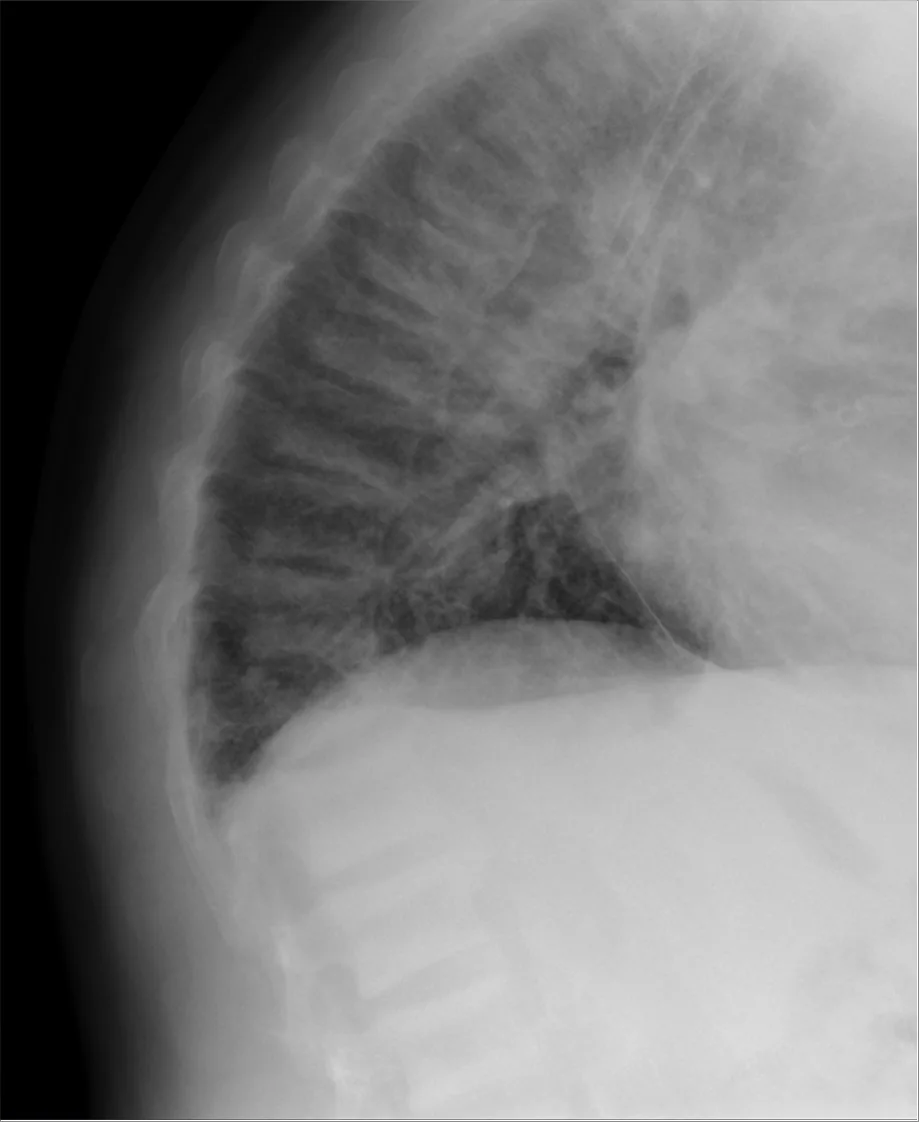 X-ray
X-rayDefinition
In secondary hyperparathyroidism (SHPT) the parathyroid glands increase PTH secretion in response to disorders outside the parathyroids, above all hypocalcemia. Unlike primary hyperparathyroidism, serum calcium is usually low or normal; once SHPT is established it may also be raised. The glands are initially regulated normally but are chronically stimulated and hyperplastic.
Long-standing SHPT can progress to tertiary hyperparathyroidism, in which PTH secretion becomes autonomous, i.e. independent of serum calcium.
Classification
- Renal SHPT: the most common form, in advanced chronic kidney disease; part of chronic kidney disease–mineral and bone disorder (CKD-MBD).
- Non-renal SHPT: vitamin D deficiency or reduced intestinal calcium absorption (e.g. in malabsorption), low calcium intake, increased renal calcium loss, or inhibition of bone resorption caused by certain drugs.
- Tertiary hyperparathyroidism: autonomous PTH secretion after long-standing SHPT, typically in end-stage kidney disease of several years' duration.
Aetiopathogenesis
In chronic kidney disease several factors interact:
- reduced renal formation of calcitriol (active vitamin D) with hypocalcemia
- phosphate retention and hyperphosphatemia
- raised fibroblast growth factor 23 (FGF-23)
SHPT can develop in moderate kidney disease with an eGFR below 60 ml/min, often before calcium or phosphate become abnormal. Marked hyperplasia reduces the sensitivity of the glands to calcium, and the calcium set point for suppressing PTH secretion rises.
In vitamin D deficiency hypocalcemia drives PTH hypersecretion. PTH increases calcium absorption, mobilisation and reabsorption but also phosphate excretion. Calcium may therefore be normal, but the hypophosphatemia impairs bone mineralisation.
Clinical features
SHPT remains clinically silent for a long time. In kidney failure the following may occur:
- osteitis fibrosa cystica with bone pain, joint symptoms and pathological fractures
- spontaneous tendon rupture
- proximal muscle weakness
- extraskeletal calcification of soft tissues and vessels
- pruritus
Renal osteodystrophy comprises increased bone turnover (hyperparathyroid bone disease) but also decreased turnover (adynamic bone disease) and osteomalacia. In vitamin D deficiency, muscle aches, muscle weakness, bone pain and a tendency to fracture predominate.
Diagnosis
- Laboratory pattern: PTH raised, calcium low or normal. Phosphate is raised in kidney disease and tends to be low in vitamin D deficiency; alkaline phosphatase is often elevated.
- Distinction from primary hyperparathyroidism in kidney disease: high phosphate with low calcium suggests secondary, high calcium with normal phosphate suggests primary hyperparathyroidism.
- Tertiary hyperparathyroidism: calcium and phosphate raised, PTH markedly elevated.
- Baseline tests: calcium, phosphate, intact PTH, 25-OH vitamin D, alkaline phosphatase and kidney function; according to KDIGO 2024 these values are measured in chronic kidney disease from stage G3a.
- Bone biopsy: the most definitive method for determining the type of renal osteodystrophy.
- Radiography: in long-standing end-stage kidney disease signs of osteitis fibrosa cystica such as subperiosteal resorption and bone cysts.
Keep learning in the app
Further reading (open access)
Cross-references
Note: Learning content for medical education – not a treatment recommendation and no substitute for diagnosis or treatment decisions in individual cases. Treatment and management are deliberately not covered on this page.