Osteomalacia and vitamin D deficiency
- Synonyms
- bone softening, rickets, vitamin D deficiency, Looser zones, hypovitaminosis D
- Specialty
- Internal medicine · Endocrinology & diabetes
- Images
- X-ray 2
- Last updated
- 10/2026 · Dr. Pascal Bafteh
Contents
Images (2)
 X-ray
X-ray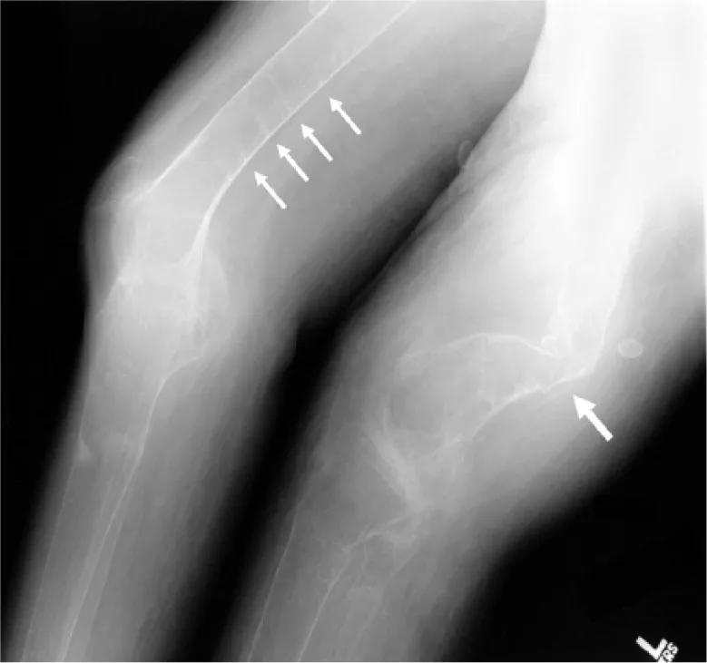 X-ray
X-rayDefinition
Osteomalacia is a disorder of mineralisation of the bone matrix in adults: the ratio of bone mineral to bone matrix is reduced. In osteoporosis, by contrast, bone mass is reduced but the mineral-to-matrix ratio is normal. In growing children, when the growth plates are also affected, the condition is called rickets.
The most common cause is severe, prolonged vitamin D deficiency or a disorder of vitamin D metabolism. Both diseases can coexist and look clinically similar.
Classification
Causes
- Vitamin D deficiency: insufficient sunlight exposure, inadequate intake, malabsorption (e.g. celiac disease).
- Abnormal vitamin D metabolism: liver disease; chronic kidney disease with reduced formation of 1,25-dihydroxyvitamin D and raised phosphate; drug-induced, e.g. by certain anticonvulsants.
- Hereditary forms: vitamin D–dependent rickets type I (autosomal recessive, defective renal conversion of 25(OH)D to 1,25-dihydroxyvitamin D) and type II (mutations of the vitamin D receptor; 1,25-dihydroxyvitamin D is abundant but ineffective).
- Phosphate deficiency: X-linked familial hypophosphatemia and other renal tubular disorders.
- Other causes: chronic metabolic acidosis, hyper- and hypoparathyroidism, inadequate calcium intake and disorders or substances that impair mineralisation of the bone matrix.
Occurrence & epidemiology
Vitamin D deficiency is common worldwide. Older people are particularly at risk – skin synthesis of vitamin D declines with age, and many get little sunlight and are undernourished –, especially care-home residents and people after a hip fracture, as well as people who stay mostly indoors or cover the whole body. Dark skin pigmentation and UV filters on the skin reduce skin synthesis. Breast milk contains little vitamin D. Osteoporosis is much more common than osteomalacia.
Aetiopathogenesis
Pathophysiology
Vitamin D3 is formed in the skin under ultraviolet B light and is obtained from food mainly from fish. The liver converts it to 25-hydroxyvitamin D, the main circulating storage form; the kidney converts this to active 1,25-dihydroxyvitamin D (calcitriol). This conversion is regulated by parathyroid hormone, calcium, phosphate and calcitriol itself. Calcitriol mainly increases intestinal absorption of calcium and phosphate and promotes bone mineralisation.
In vitamin D deficiency, hypocalcemia causes secondary hyperparathyroidism. This raises calcium through bone mobilisation and renal reabsorption but increases phosphate excretion. Serum calcium may therefore be normal, but hypophosphatemia impairs adequate mineralisation.
Clinical features
- Adults: muscle aches, muscle weakness and bone pain; increased fracture tendency, recurrent rib or unusual fractures; in older people hip fractures after minimal trauma.
- Hypocalcemic tetany: paraesthesia of the lips, tongue and fingers, carpopedal and facial spasm, in severe cases seizures.
- Rickets in infants and toddlers: craniotabes (softening of the skull), delayed sitting, crawling and walking, delayed fontanelle closure, frontal bossing, "rachitic rosary" at the costochondral junctions, enlarged wrists and ankles, kyphoscoliosis.
- Rickets in older children and adolescents: painful walking, bowlegs or knock-knees, flattened pelvis.
- Maternal vitamin D deficiency causes deficiency in the fetus and can cause neonatal tetany.
Histology
The hallmark is unmineralised osteoid with reduced mineral content. Definitive distinction from osteoporosis is possible with a tetracycline-labelled bone biopsy; this is rarely needed, e.g. with persistently very low vitamin D or unexplained fragility fractures in young people.
Diagnosis
- 25-hydroxyvitamin D (D2 + D3): the best measure of vitamin D stores and for diagnosing deficiency.
- Associated laboratory findings: calcium low or normal because of secondary hyperparathyroidism, phosphate usually low, alkaline phosphatase usually raised, parathyroid hormone normal or raised; urinary calcium low (except with acidosis).
- 1,25-dihydroxyvitamin D: markedly low to undetectable in severe deficiency; in vitamin D–dependent rickets type I normal 25(OH)D, low 1,25-dihydroxyvitamin D and calcium, normal or low phosphate.
- Radiography in adults: demineralisation especially of the spine, pelvis and legs; ribbon-like lucencies in the cortex (Looser zones, pseudofractures).
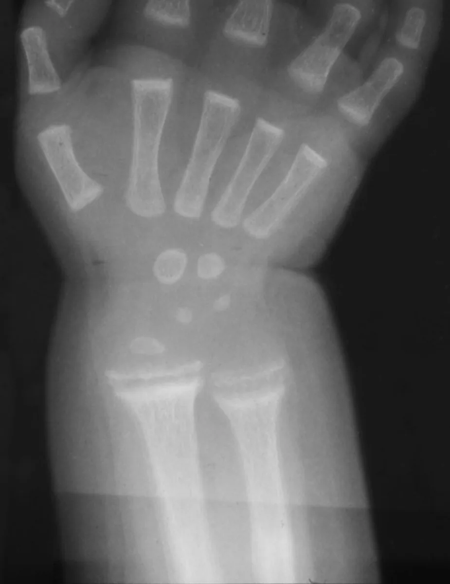
- Radiography in rickets: most evident at the distal radius and ulna; the metaphyses lose their sharp outline and become cupped with fringy rarefaction; the distance to the metacarpals appears increased. Bone changes precede clinical signs.
- Distinction from osteoporosis: both can look similar on imaging; osteomalacia is suggested by bone pain, unusual fractures and persistently very low vitamin D.
Keep learning in the app
Further reading (open access)
Cross-references
Note: Learning content for medical education – not a treatment recommendation and no substitute for diagnosis or treatment decisions in individual cases. Treatment and management are deliberately not covered on this page.