Multiple endocrine neoplasia (MEN 1 and MEN 2)
- Synonyms
- MEN, MEN1, MEN2, Wermer syndrome, Sipple syndrome, multiple endocrine neoplasia type 2
- Specialty
- Internal medicine · Endocrinology & diabetes
- Images
- Histology 1 · Ultrasound 1
- Last updated
- 10/2026 · Dr. Pascal Bafteh
Contents
Images (2)
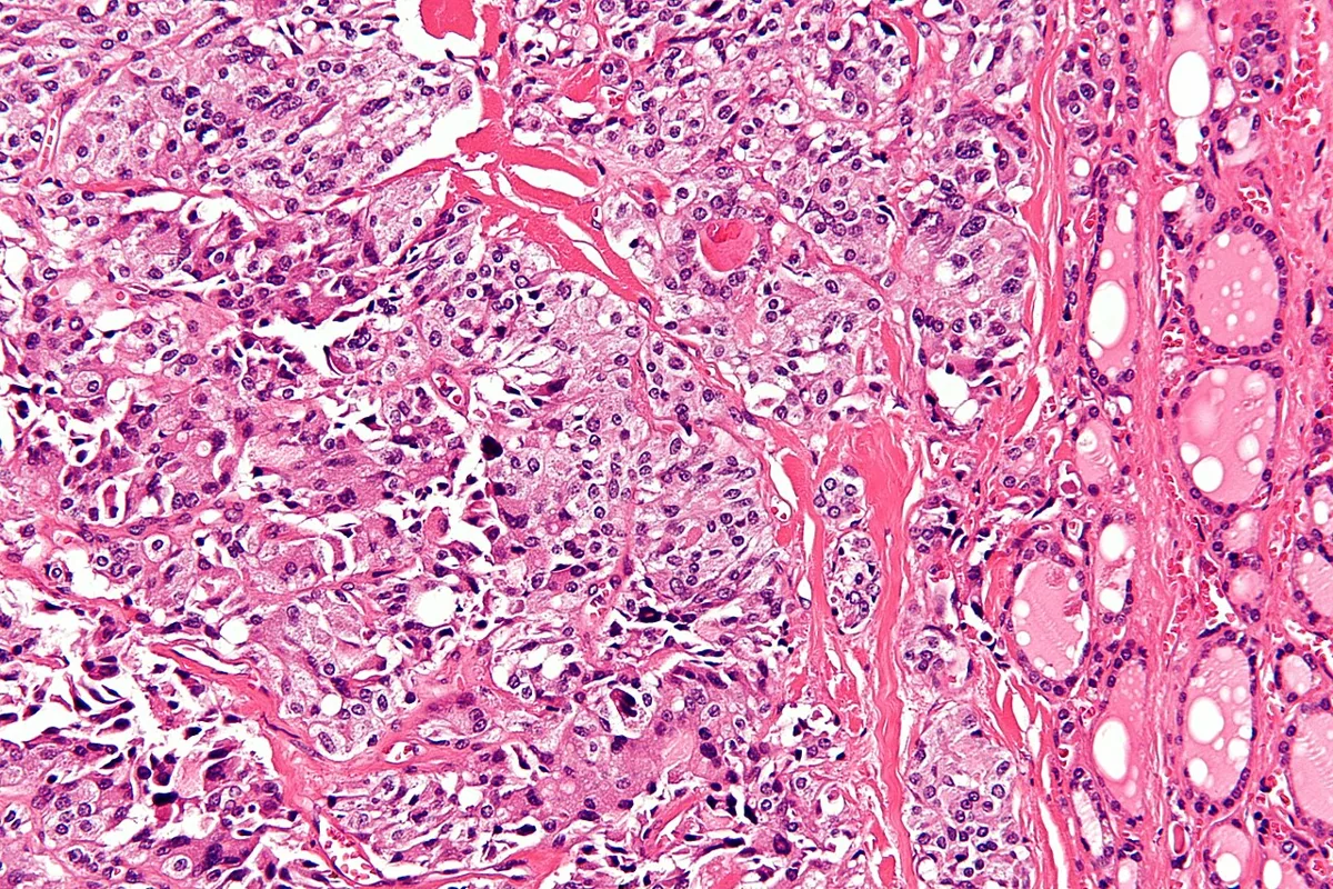 Histology
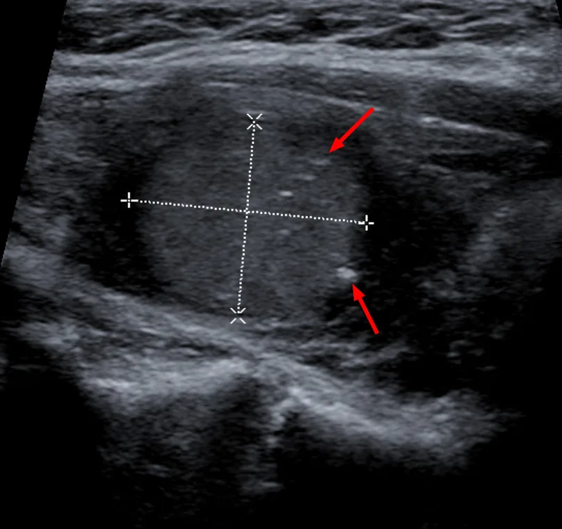 Ultrasound
Definition
Multiple endocrine neoplasias (MEN) are autosomal dominant inherited syndromes in which hyperplasia or tumors develop in several endocrine glands, simultaneously or sequentially.
- MEN 1: parathyroids, endocrine pancreas or duodenum and pituitary; caused by inactivating mutations of the MEN1 gene, which encodes the tumor suppressor protein menin.
- MEN 2A: medullary thyroid carcinoma, phaeochromocytoma and primary hyperparathyroidism; familial medullary thyroid carcinoma is considered a distinct variant.
- MEN 2B: medullary thyroid carcinoma, phaeochromocytoma, mucosal neuromas, intestinal ganglioneuromatosis and a marfanoid habitus – without hyperparathyroidism.
MEN 2A, MEN 2B and familial medullary carcinoma are caused by activating mutations of the RET proto-oncogene on chromosome 10, which encodes a receptor tyrosine kinase.
Classification
MEN 1 – manifestations
- Primary hyperparathyroidism: in 95 % or more; diffuse, often asymmetric hyperplasia of several glands is typical.
- Pancreatic or duodenal neuroendocrine tumors: in 30–90 %; often multicentric. The most common functioning tumor is gastrinoma, followed by insulinoma; about one third of patients have non-functioning tumors. About 30 % are already metastatic at diagnosis.
- Pituitary tumors: in 15–42 %; of these 25–90 % are prolactinomas, about 25 % secrete GH or GH and prolactin, about 3 % secrete ACTH, and most of the rest are non-functioning.
- Other manifestations: foregut carcinoids (thymus, lung, stomach) in 5–15 %, thymic carcinoids more often in men; adrenal adenomas in up to 33 %; lipomas, angiofibromas, collagenomas, meningiomas and ependymomas; increased risk of breast cancer.
MEN 2A and MEN 2B – manifestations
- MEN 2A: medullary thyroid carcinoma in almost all patients; phaeochromocytoma in 40–50 % of affected family members, bilateral and multicentric in more than half; hyperparathyroidism in 10–20 %, often involving several glands; cutaneous lichen amyloidosis in some families; Hirschsprung disease in 2–5 %.
- MEN 2B: about 95 % carry the same point mutation in the RET protein (amino acid 918), more than half as new mutations. About 50 % have the complete syndrome of mucosal neuromas, phaeochromocytoma and medullary carcinoma; the medullary carcinoma is particularly aggressive and almost always present by the first year of life.
Occurrence & epidemiology
MEN 1: onset between 4 and 81 years, peak incidence in the third to fifth decades; both sexes equally affected. More than 50 % of mutation carriers develop the disease by age 40 and more than 95 % by age 80. About 40 % have tumors in all three main organs. MEN 2: Of all medullary thyroid carcinomas about 25 % are familial; up to about 7 % of apparently sporadic cases carry a RET mutation.
Clinical features
MEN 1
- Hyperparathyroidism: usually asymptomatic hypercalcemia; about 25 % have kidney stones or nephrocalcinosis.
- Gastrinoma: up to 80 % of patients with MEN 1 have multiple peptic ulcers due to acid hypersecretion or asymptomatically raised gastrin.
- Insulinoma: fasting hypoglycemia, often before the age of 40; tumors are small and often multiple.
- Other pancreatic tumors: watery diarrhea with hypokalemia and achlorhydria (WDHA syndrome); less often ectopic ACTH or CRH (Cushing's syndrome) or GHRH (acromegaly).
- Pituitary tumors: galactorrhoea, acromegaly, Cushing's disease; mass effects with visual disturbance, headache and hypopituitarism.
MEN 2A and MEN 2B
- Medullary thyroid carcinoma: usually develops in childhood from C-cell hyperplasia and is often multicentric.
- Phaeochromocytoma: in MEN 2 produces disproportionately more adrenaline; hypertension is more often paroxysmal than sustained, and hypertensive crises are typical; many patients, however, are asymptomatic.
- Hyperparathyroidism (MEN 2A only): hypercalcemia, kidney stones, nephrocalcinosis.
- Cutaneous lichen amyloidosis (MEN 2A): pruritic, scaly papules between the shoulder blades or on extensor surfaces.
- MEN 2B: glistening bumps (neuromas) on the lips, tongue and buccal mucosa, often the earliest sign; neuromas of the eyelids, conjunctivae and corneas, thickened eyelids, everted lips; altered bowel motility with constipation, diarrhea and occasionally megacolon; marfanoid habitus and skeletal deformities.
Diagnosis
- Suspected MEN 1: with tumors of the parathyroids, pancreas or pituitary, especially with a family history; with primary hyperparathyroidism before the age of 30.
- Laboratory tests in MEN 1: calcium, intact PTH, gastrin and prolactin. Gastrinoma: raised basal gastrin with a paradoxical rise in the secretin test; insulinoma: fasting hypoglycemia with a raised insulin level; acromegaly: raised IGF-1 and absent GH suppression in the glucose tolerance test.
- Imaging in MEN 1: ultrasound, endoscopic ultrasound, CT and MRI; gallium-68 DOTATATE PET/CT was about three times more sensitive than older scintigraphic and CT methods for pancreatic and duodenal tumors and helps to detect thymic and lung carcinoids.
- Laboratory tests in MEN 2: serum calcitonin (medullary carcinoma), calcium, 24-hour urinary calcium and PTH, plasma free metanephrines or urinary catecholamines and metanephrines.
- Imaging in MEN 2: neck ultrasound, complemented by CT, MRI or scintigraphy; CT or MRI to localise phaeochromocytomas and detect bilateral tumors.
- Genetics: panel testing when MEN 1 is suspected (including genes for MEN 4 and other forms of familial hyperparathyroidism); RET testing in all patients with medullary thyroid carcinoma. The specific RET mutation predicts aggressiveness and associated manifestations. First-degree relatives are offered genetic testing.
Keep learning in the app
Further reading (open access)
Cross-references
Note: Learning content for medical education – not a treatment recommendation and no substitute for diagnosis or treatment decisions in individual cases. Treatment and management are deliberately not covered on this page.