Lupus nephritis
Board exam relevance: in 1 of 105 exam reports · rank 181- Synonyms
- SLE nephritis, lupus glomerulonephritis, kidney involvement in lupus
- Specialty
- Internal medicine · Nephrology
- Images
- Histology 1
- Last updated
- 10/2026 · Dr. Pascal Bafteh
Contents
Images (1)
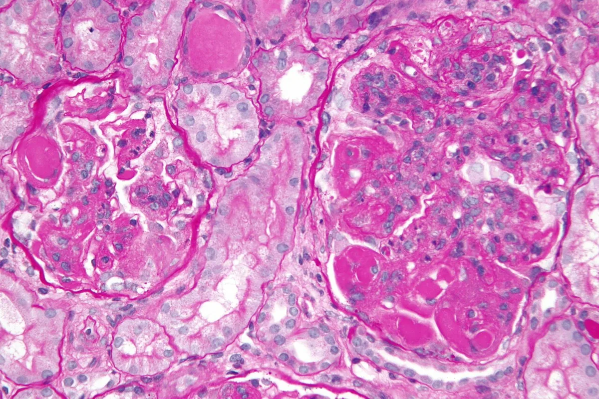 Histology
Definition
Lupus nephritis is glomerulonephritis caused by systemic lupus erythematosus (SLE). It results from deposition of immune complexes composed of nuclear antigens (especially DNA) and complement-fixing autoantibodies in the glomeruli.
Classification
ISN/RPS classification
The histologic ISN/RPS classification (2003, revised 2018) distinguishes six classes:
- Class I – minimal mesangial: normal on light microscopy, immune complexes only on immunofluorescence or electron microscopy
- Class II – mesangial proliferative: immune complexes in the mesangium only, mesangial hypercellularity; possibly microscopic hematuria or proteinuria
- Class III – focal: endocapillary and extracapillary proliferation in < 50% of glomeruli, usually segmental
- Class IV – diffuse: proliferation in ≥ 50% of glomeruli; most common form, often with hypertension, nephrotic syndrome and elevated creatinine
- Class V – membranous: thickened basement membrane with subepithelial and intramembranous immune complexes; usually nephrotic syndrome with normal or slightly elevated creatinine
- Class VI – advanced sclerosing: ≥ 90% of glomeruli globally sclerosed without residual activity; bland sediment and kidney failure
Overlap and transitions between classes are common.
Occurrence & epidemiology
Lupus nephritis occurs in 20–60% of people with SLE. In the United States, African American patients are disproportionately affected.
Clinical features
The other manifestations of SLE are usually in the foreground. Renal findings include hematuria, proteinuria (often nephrotic-range, ≥ 3 g/day), edema, hypertension and, in advanced stages, kidney failure. Antiphospholipid syndrome nephropathy with microthrombi and ischemic cortical atrophy may additionally be present in up to one third of patients with SLE.
Histology
Depending on the class, immune complexes lie subendothelially, intramembranously, subepithelially or in the mesangium. Immunofluorescence shows complement and IgG, IgA and IgM in varying proportions; epithelial cell proliferation may form crescents.
Diagnosis
- In everyone with SLE: urinalysis with sediment (hematuria, red blood cell casts), protein-to-creatinine ratio and serum creatinine
- Anti-dsDNA antibodies and complement C3 and C4: high anti-dsDNA titers and low complement suggest active lupus nephritis.
- Kidney biopsy if findings are abnormal, especially with declining GFR or confirmed proteinuria: confirmation of the diagnosis and determination of the ISN/RPS class
- Lupus nephritis is also considered in unexplained hypertension, elevated creatinine or abnormal urinalysis with clinical features of lupus.
Keep learning in the app
Further reading (open access)
Cross-references
More topics: Nephrology
- Acute kidney injury
- Nephrotic syndrome
- Chronic kidney disease
- Nephritic syndrome and glomerulonephritis
- Acute interstitial nephritis
- Goodpasture syndrome (anti-GBM disease)
- Membranous nephropathy
- Renal artery stenosis
- Diabetic kidney disease
- Urinary tract infection and cystitis
- Acute pyelonephritis
- Autosomal dominant polycystic kidney disease (ADPKD)
Note: Learning content for medical education – not a treatment recommendation and no substitute for diagnosis or treatment decisions in individual cases. Treatment and management are deliberately not covered on this page.