Membranous nephropathy
Board exam relevance: in 3 of 105 exam reports · rank 111- Synonyms
- membranous glomerulonephritis, MGN, MN, PLA2R-associated nephropathy
- Specialty
- Internal medicine · Nephrology
- Images
- Histology 1
- Last updated
- 10/2026 · Dr. Pascal Bafteh
Contents
Images (1)
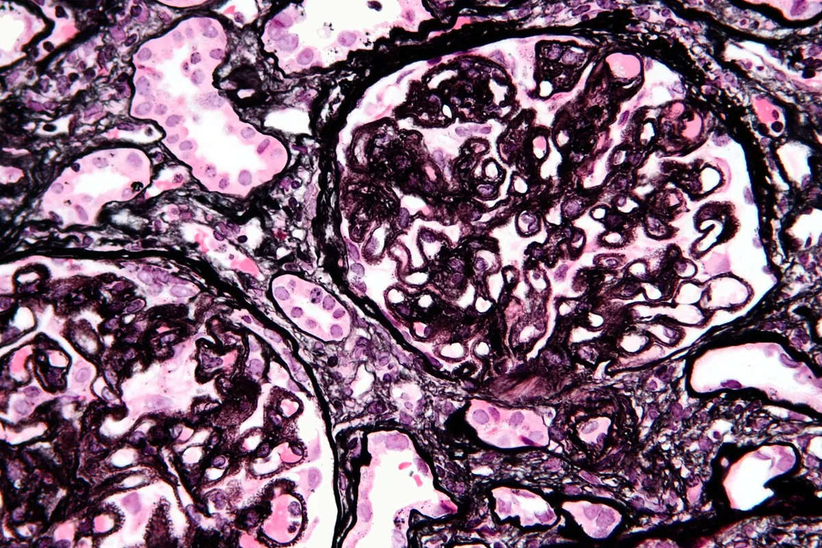 Histology
Definition
Membranous nephropathy (membranous glomerulonephritis) is characterized by deposition of immune complexes on the outer side of the glomerular basement membrane (subepithelial) with thickening of the basement membrane. It mainly affects adults, in whom it is a common cause of nephrotic syndrome.
Aetiopathogenesis
In the primary form, the M-type phospholipase A2 receptor (PLA2R) on podocytes is the main antigen of the deposited immune complexes.
Secondary forms occur with:
- cancer (in 6–15% of patients), e.g. carcinomas of the lung, prostate, colon, stomach, breast and kidney, lymphomas, chronic lymphocytic leukemia, melanoma
- infections: hepatitis B and C, syphilis, HIV, malaria, schistosomiasis
- autoimmune diseases: systemic lupus erythematosus, thyroiditis
- certain drugs, e.g. NSAIDs
In children the disease is rare and usually secondary, e.g. to chronic hepatitis B, lupus or autoimmune thyroid disease.
Clinical features
Typical is a nephrotic syndrome of insidious onset with edema; nephrotic-range proteinuria is present in about 80%. The urine sediment is usually bland, occasionally with microscopic hematuria and hypertension; kidney function is often normal initially.
Renal vein thrombosis is more frequent in membranous nephropathy than in other glomerulopathies. It is often silent but may cause flank pain, hematuria and hypertension and may lead to pulmonary embolism.
Histology
Electron microscopy shows subepithelial electron-dense deposits, between which the basement membrane grows outward as "spikes" (silver stain). Later, the deposits lie within the markedly thickened basement membrane. Immunofluorescence shows diffuse granular IgG deposition along the capillary walls without cellular proliferation or necrosis. In the primary PLA2R-positive form IgG4 predominates, in cancer-associated forms IgG1 and IgG2.
Diagnosis
- basic workup of nephrotic syndrome: protein-to-creatinine ratio, serum albumin, lipids, creatinine and eGFR
- serum anti-PLA2R antibodies to distinguish primary from secondary forms
- kidney biopsy to confirm the diagnosis
- search for secondary causes: age-appropriate cancer screening (especially with weight loss, anemia or older age), hepatitis B and C serology, antinuclear antibodies, drug history
Keep learning in the app
Further reading (open access)
Cross-references
More topics: Nephrology
- Acute kidney injury
- Nephrotic syndrome
- Chronic kidney disease
- Nephritic syndrome and glomerulonephritis
- Acute interstitial nephritis
- Goodpasture syndrome (anti-GBM disease)
- Renal artery stenosis
- Diabetic kidney disease
- Urinary tract infection and cystitis
- Acute pyelonephritis
- Autosomal dominant polycystic kidney disease (ADPKD)
- Hantavirus infection (nephropathia epidemica)
Note: Learning content for medical education – not a treatment recommendation and no substitute for diagnosis or treatment decisions in individual cases. Treatment and management are deliberately not covered on this page.