Nephritic syndrome and glomerulonephritis
Board exam relevance: in 4 of 105 exam reports · rank 90- Synonyms
- glomerulonephritis, GN, kidney inflammation, poststreptococcal glomerulonephritis, postinfectious glomerulonephritis
- Specialty
- Internal medicine · Nephrology
- Images
- Blood smear & cytology 1 · Histology 1
- Last updated
- 10/2026 · Dr. Pascal Bafteh
Contents
Images (2)
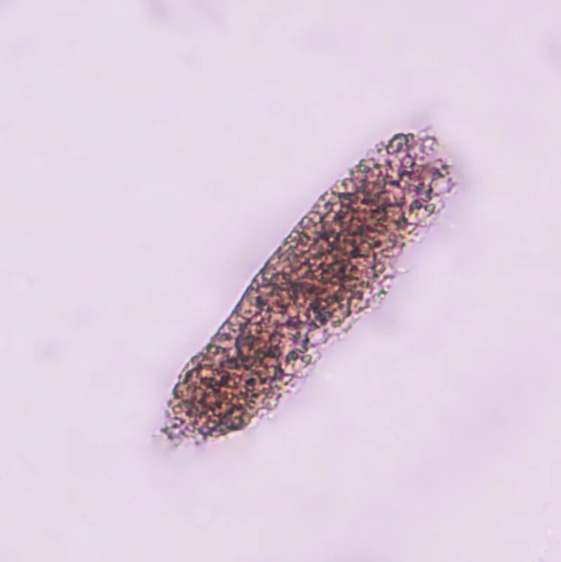 Blood smear & cytology
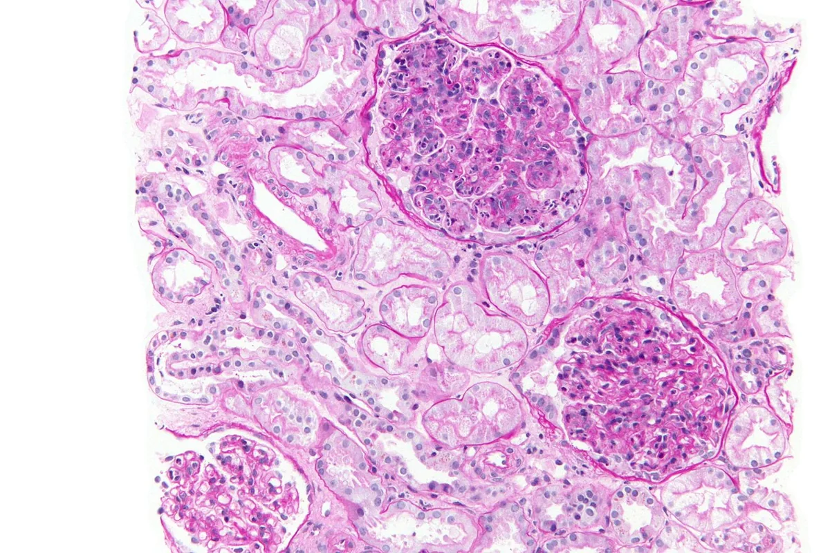 Histology
Definition
Nephritic syndrome is the clinical expression of glomerular inflammation (glomerulonephritis). Its hallmarks are hematuria with usually dysmorphic red blood cells and often red blood cell casts, together with variable proteinuria. Edema, hypertension, a rise in creatinine and oliguria are often present as well.
Classification
Types
- Acute glomerulonephritis: creatinine rises over days to weeks; the prototype is postinfectious glomerulonephritis
- Rapidly progressive glomerulonephritis: kidney failure within weeks to months with crescent formation
- Chronic glomerulonephritis: slow course over years with mild to moderate proteinuria, e.g. IgA nephropathy, Alport syndrome
- In addition, primary (kidney-limited) and secondary forms due to infections or systemic diseases are distinguished.
Aetiopathogenesis
- Primary: IgA nephropathy, membranoproliferative glomerulonephritis, idiopathic crescentic glomerulonephritis
- Infection-related: group A streptococci, staphylococci (especially endocarditis), hepatitis B and C, visceral abscesses, malaria
- Systemic diseases: systemic lupus erythematosus, IgA vasculitis, ANCA-associated vasculitides, anti-GBM disease, cryoglobulinemia, hemolytic uremic syndrome
- Hereditary: Alport syndrome, thin basement membrane disease
Postinfectious glomerulonephritis is the most common acute glomerulonephritis in children aged 3 to 15 years. It typically follows pharyngitis or impetigo due to nephritogenic group A streptococci after 6–21 days (up to 6 weeks). Microbial antigens and immune complexes deposit in the glomeruli and activate mainly the alternative complement pathway.
Clinical features
The spectrum ranges from asymptomatic microscopic hematuria to the full picture with cola- or smoke-colored urine, oliguria, edema, hypertension and acute kidney injury. Systemic diseases additionally present with skin lesions, joint symptoms, hemoptysis or constitutional symptoms. Fever is unusual in the postinfectious form and suggests persistent infection.
Histology
In postinfectious glomerulonephritis, the glomeruli are enlarged and hypercellular, with initial neutrophilic infiltration, proliferation of endothelial and mesangial cells and subepithelial electron-dense deposits; crescents form in severe cases.
Diagnosis
- Urine sediment: glomerular red cells are smaller and dysmorphic (spicules, folding, blebs); red blood cell casts and dysmorphic red cells indicate glomerulonephritis or vasculitis.
- Quantify proteinuria (protein-to-creatinine ratio); in the postinfectious form usually 0.2–2, occasionally in the nephrotic range
- Kidney function: creatinine, urea, electrolytes
- Complement: low C3 (and CH50) in postinfectious glomerulonephritis, usually normalizing within 6–8 weeks; low C3 and C4 also in active lupus nephritis
- Serology: antistreptolysin O, anti-DNase B, antinuclear and anti-dsDNA antibodies, ANCA, anti-GBM antibodies, hepatitis serology
- Kidney biopsy with light microscopy, immunofluorescence and electron microscopy to determine the cause
Keep learning in the app
Further reading (open access)
Cross-references
More topics: Nephrology
- Acute kidney injury
- Nephrotic syndrome
- Chronic kidney disease
- Acute interstitial nephritis
- Goodpasture syndrome (anti-GBM disease)
- Membranous nephropathy
- Renal artery stenosis
- Diabetic kidney disease
- Urinary tract infection and cystitis
- Acute pyelonephritis
- Autosomal dominant polycystic kidney disease (ADPKD)
- Hantavirus infection (nephropathia epidemica)
Note: Learning content for medical education – not a treatment recommendation and no substitute for diagnosis or treatment decisions in individual cases. Treatment and management are deliberately not covered on this page.