Diabetic kidney disease
Board exam relevance: in 2 of 105 exam reports · rank 142- Synonyms
- DKD, Kimmelstiel-Wilson disease, diabetic glomerulosclerosis
- Specialty
- Internal medicine · Nephrology
- Images
- Histology 1
- Last updated
- 10/2026 · Dr. Pascal Bafteh
Contents
Images (1)
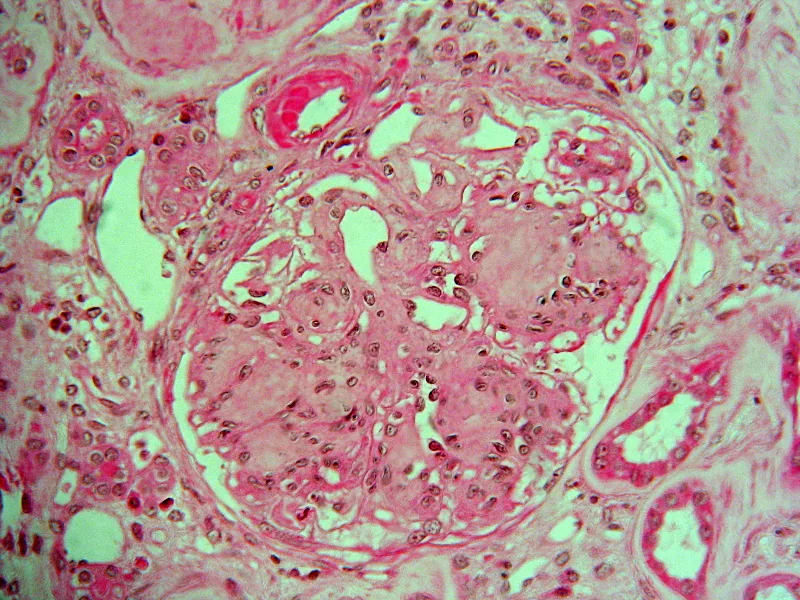 Histology
Definition
Diabetic nephropathy (diabetic kidney disease) is glomerular sclerosis and fibrosis caused by the metabolic and hemodynamic changes of diabetes mellitus. It manifests as slowly progressive albuminuria with worsening hypertension and progressive chronic kidney disease.
Occurrence & epidemiology
Diabetic nephropathy is the most common cause of kidney failure worldwide and the most common cause of nephrotic syndrome in adults. Because type 2 diabetes often remains unrecognized for years, nephropathy may already be present at diagnosis; about 25% of people with type 2 diabetes have microalbuminuria 10 years after diagnosis.
Aetiopathogenesis
Risk factors and pathogenesis
Risk factors are the duration and degree of hyperglycemia, arterial hypertension, dyslipidemia, smoking, family history and genetic factors such as polymorphisms of the renin-angiotensin-aldosterone system or a reduced number of glomeruli.
The process begins with microangiopathy. Glycation of glomerular proteins, cytokines such as TGF-β, proliferation of mesangial cells and matrix and altered glomerular hemodynamics lead to thickening of the basement membrane and sclerosis. An early functional abnormality is glomerular hyperfiltration.
Clinical features
Typical course over years: glomerular hyperfiltration, then moderately increased albuminuria (30–300 mg/24 h, formerly microalbuminuria), later severely increased albuminuria (> 300 mg/24 h) and often nephrotic syndrome, followed several years later by kidney failure. Early stages are asymptomatic; persistent microalbuminuria is the earliest warning sign.
In the course, most patients develop hypertension and edema. Uremic symptoms often appear at a higher GFR than in other kidney diseases. Papillary necrosis, type 4 renal tubular acidosis and urinary tract infections occur more often.
Histology
Thickened glomerular basement membrane, diffuse or nodular intercapillary glomerulosclerosis (the nodular form is called the Kimmelstiel-Wilson lesion), marked hyalinosis of afferent and efferent arterioles, and interstitial fibrosis and tubular atrophy. The extent of mesangial expansion correlates with progression to kidney failure.
Diagnosis
- Albumin-to-creatinine ratio in a spot urine, ideally mid-morning: a value ≥ 30 mg/g in at least 2 of 3 samples within 3–6 months indicates moderately increased albuminuria, provided infection or exercise has been excluded as the cause.
- If the dipstick is already positive for protein, severely increased albuminuria is usually present.
- eGFR and urine sediment; on ultrasound the kidneys are usually normal-sized or enlarged.
- The presence of diabetic retinopathy supports the diagnosis as a sign of microangiopathy.
The diagnosis is usually clinical. Another kidney disease is considered in case of heavy proteinuria with a short duration of diabetes, absent retinopathy, rapid onset of heavy proteinuria, gross hematuria, red blood cell casts, rapid decline in GFR or small kidneys; a kidney biopsy then clarifies the cause.
Keep learning in the app
Further reading (open access)
Cross-references
More topics: Nephrology
- Acute kidney injury
- Nephrotic syndrome
- Chronic kidney disease
- Nephritic syndrome and glomerulonephritis
- Acute interstitial nephritis
- Goodpasture syndrome (anti-GBM disease)
- Membranous nephropathy
- Renal artery stenosis
- Urinary tract infection and cystitis
- Acute pyelonephritis
- Autosomal dominant polycystic kidney disease (ADPKD)
- Hantavirus infection (nephropathia epidemica)
Note: Learning content for medical education – not a treatment recommendation and no substitute for diagnosis or treatment decisions in individual cases. Treatment and management are deliberately not covered on this page.