Nephrotic syndrome
Board exam relevance: in 8 of 105 exam reports · rank 39- Synonyms
- nephrosis, heavy proteinuria, protein-losing kidney disease
- Specialty
- Internal medicine · Nephrology
- Images
- Blood smear & cytology 1
- Last updated
- 10/2026 · Dr. Pascal Bafteh
Contents
Images (1)
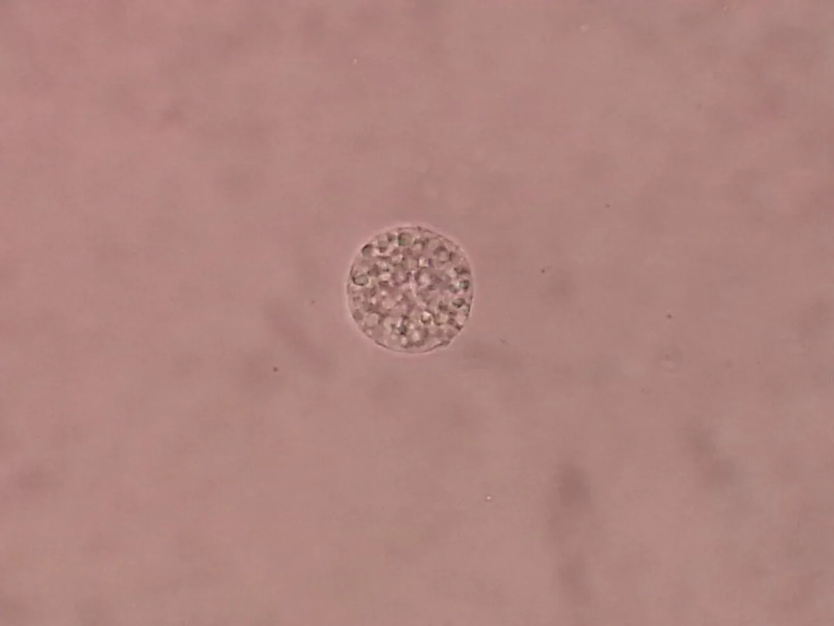 Blood smear & cytology
Definition
Nephrotic syndrome is characterized by heavy glomerular proteinuria in the nephrotic range (> 3.5 g/day; some definitions use ≥ 3 g/day), hypoalbuminemia and edema; hyperlipidemia and lipiduria are usually also present. It is not a single disease but a syndrome caused by various glomerulopathies.
Occurrence & epidemiology
Nephrotic syndrome occurs at any age but is more common in children (mean age at diagnosis 5–6 years, mostly minimal change disease). Below 8 years, boys are affected more often than girls; at older ages both sexes equally. Congenital forms present in the first year of life.
Aetiopathogenesis
- Primary causes: minimal change disease, focal segmental glomerulosclerosis, membranous nephropathy; less often membranoproliferative glomerulonephritis or IgA nephropathy
- Secondary causes (more common in adults): diabetic nephropathy, systemic lupus erythematosus, amyloidosis, preeclampsia, cancer (carcinomas, lymphomas, myeloma), infections (hepatitis B and C, HIV, syphilis, malaria), drugs and substances (e.g. NSAIDs, heroin), severe obesity, genetic syndromes
Pathophysiologically, the endothelial cells, glomerular basement membrane or podocytes that normally retain proteins by size and charge are damaged. Besides albumin, antibodies, opsonins, antithrombin III, transferrin and binding proteins for hormones and vitamin D are lost in the urine, which explains the complications.
Clinical features
Symptoms and complications
Leading symptoms are edema (peripheral edema, plus ascites and pleural effusions), frothy urine, anorexia and malaise. Effusions may cause dyspnea, joint symptoms or abdominal pain; white transverse lines on the fingernails (Muehrcke lines) may appear.
- Thromboembolism, especially renal vein thrombosis and pulmonary embolism (loss of antithrombin III, increased hepatic synthesis of clotting factors)
- Infections such as cellulitis and spontaneous bacterial peritonitis (loss of antibodies and opsonins)
- Dyslipidemia with increased atherosclerotic risk, hypertension, anemia, vitamin D deficiency and chronic kidney disease
Diagnosis
- Proteinuria: protein-to-creatinine ratio in a spot urine or 24-hour urine collection (normal < 150 mg/24 h)
- Urine sediment: lipiduria with oval fat bodies and fatty casts; cholesterol shows a Maltese cross pattern under polarized light
- Blood: serum albumin often < 2.5 g/dL (25 g/L), elevated total cholesterol and triglycerides, creatinine and urea depending on kidney function
- Search for secondary causes depending on the clinical context: HbA1c, antinuclear antibodies, hepatitis B and C serology, HIV and syphilis serology, serum and urine protein electrophoresis, cryoglobulins, complement C3 and C4
- Kidney biopsy: in adults with an unclear cause to diagnose the underlying glomerulopathy; in children with a typical presentation, minimal change disease is usually presumed
Keep learning in the app
Further reading (open access)
Cross-references
More topics: Nephrology
- Acute kidney injury
- Chronic kidney disease
- Nephritic syndrome and glomerulonephritis
- Acute interstitial nephritis
- Goodpasture syndrome (anti-GBM disease)
- Membranous nephropathy
- Renal artery stenosis
- Diabetic kidney disease
- Urinary tract infection and cystitis
- Acute pyelonephritis
- Autosomal dominant polycystic kidney disease (ADPKD)
- Hantavirus infection (nephropathia epidemica)
Note: Learning content for medical education – not a treatment recommendation and no substitute for diagnosis or treatment decisions in individual cases. Treatment and management are deliberately not covered on this page.