Acute kidney injury
Board exam relevance: in 9 of 105 exam reports · rank 32- Synonyms
- acute renal failure, AKI, kidney failure, acute tubular necrosis
- Specialty
- Internal medicine · Nephrology
- Images
- Blood smear & cytology 2 · Histology 1 · Gross specimen 1
- Last updated
- 10/2026 · Dr. Pascal Bafteh
Contents
Images (4)
 Blood smear & cytology
 Histology
 Blood smear & cytology
 Gross specimen
Definition
Acute kidney injury (AKI; formerly acute renal failure) is a rapid decline in kidney function over days to weeks with a rise in blood creatinine and urea (azotemia), with or without a fall in urine output. According to KDIGO, AKI is present in case of:
- a rise in serum creatinine by ≥ 0.3 mg/dL (26.5 µmol/L) within 48 hours, or
- a rise to ≥ 1.5 times baseline within the prior 7 days, or
- urine output < 0.5 mL/kg/h for 6 hours
Classification
KDIGO stages and types
- Stage 1: creatinine 1.5 to 1.9 times baseline or a rise of ≥ 0.3 mg/dL; urine < 0.5 mL/kg/h for 6–12 hours
- Stage 2: creatinine 2.0 to 2.9 times baseline; urine < 0.5 mL/kg/h for ≥ 12 hours
- Stage 3: creatinine ≥ 3 times baseline or ≥ 4.0 mg/dL (353.6 µmol/L); urine < 0.3 mL/kg/h for ≥ 24 hours or anuria for ≥ 12 hours
By cause, prerenal, intrinsic renal and postrenal AKI are distinguished; by urine volume, oliguric (< 500 mL/day) and nonoliguric courses.
Occurrence & epidemiology
AKI is a common organ dysfunction in hospital inpatients, particularly in sepsis, major trauma and circulatory failure. Prerenal AKI and acute tubular injury are the most common causes in this setting.
Aetiopathogenesis
Causes
- Prerenal (reduced renal perfusion of intact tissue): volume depletion (vomiting, diarrhea, hemorrhage, burns), heart failure and cardiogenic shock, sepsis, decompensated liver cirrhosis, drugs such as NSAIDs. The kidney retains sodium and water; sustained hypoperfusion progresses to ischemic tubular injury.
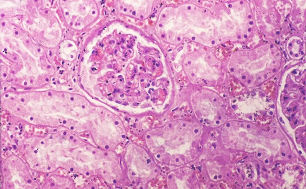
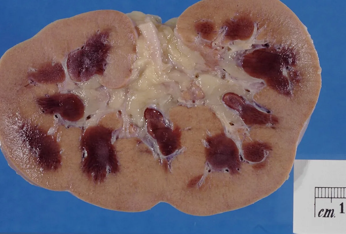
- Intrinsic renal: acute tubular necrosis (mostly ischemic; toxic e.g. due to aminoglycosides, iodinated contrast media, myoglobin in rhabdomyolysis, hemoglobin in hemolysis), glomerulonephritis (ANCA-associated, anti-GBM, immune complex), acute interstitial nephritis and vascular disorders (atheroembolism, thrombotic microangiopathy, malignant hypertension).
- Postrenal: obstruction between the tubules and the urethra, including microscopic obstruction within the tubules by precipitating crystals or proteins (e.g. uric acid, myeloma light chains); back pressure reduces glomerular filtration. Ureteral obstruction causes AKI only if bilateral or in a single kidney. The most common cause of sudden urinary retention in men is prostatic enlargement; other causes are calculi, tumors and neurogenic bladder disorders.
Clinical features
Symptoms
Symptoms of the underlying illness often predominate. Early signs are falling urine output, weight gain and edema; as uremia progresses, anorexia, nausea, vomiting, weakness, myoclonic jerks, confusion, seizures and finally coma follow. Dark, tea-colored urine suggests glomerulonephritis or myoglobinuria, a palpable bladder suggests bladder outlet obstruction.
Course of urine output
Typical is a prodromal phase with usually normal urine output, an oliguric phase with 50–500 mL of urine per day (many patients are never oliguric) and a polyuric recovery phase in which sodium wasting and polyuria may persist for days to weeks.
Complications
- hyperkalemia with cardiac arrhythmias
- fluid overload up to pulmonary edema
- metabolic acidosis, hyperphosphatemia, hypocalcemia
- uremic pericarditis, encephalopathy and bleeding tendency
Diagnosis
Laboratory tests
- Creatinine and urea rise day by day, creatinine by up to 2 mg/dL (180 µmol/L) daily. GFR estimating equations are unreliable while creatinine is unstable.
- Electrolytes and blood gases: hyperkalemia, hyperphosphatemia, hypocalcemia, usually moderate hyponatremia (125–135 mmol/L), metabolic acidosis with bicarbonate usually 15–20 mmol/L
- Blood count: normochromic, normocytic anemia (hematocrit typically 25–30%)
- Depending on suspicion: creatine kinase and myoglobin (rhabdomyolysis), uric acid (tumor lysis), serum protein electrophoresis (myeloma)
Urine findings
In oliguria, urinary indices distinguish prerenal AKI from tubular injury:
- fractional excretion of sodium (FENa) < 1% prerenal, > 1% in tubular injury
- urine sodium < 10 mmol/L prerenal, > 40 mmol/L in tubular injury
- urine-to-plasma osmolality ratio > 1.5 prerenal, 1–1.5 in tubular injury
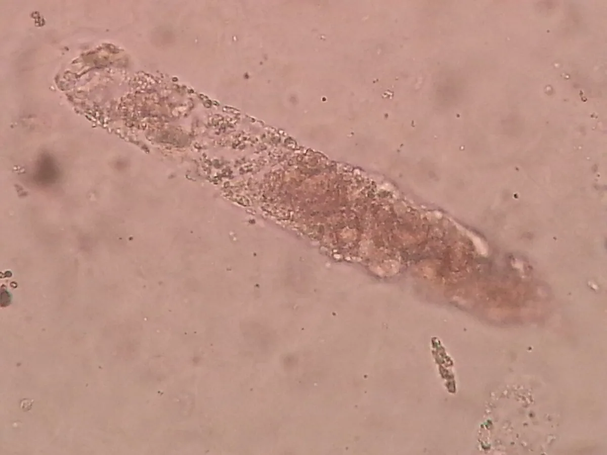
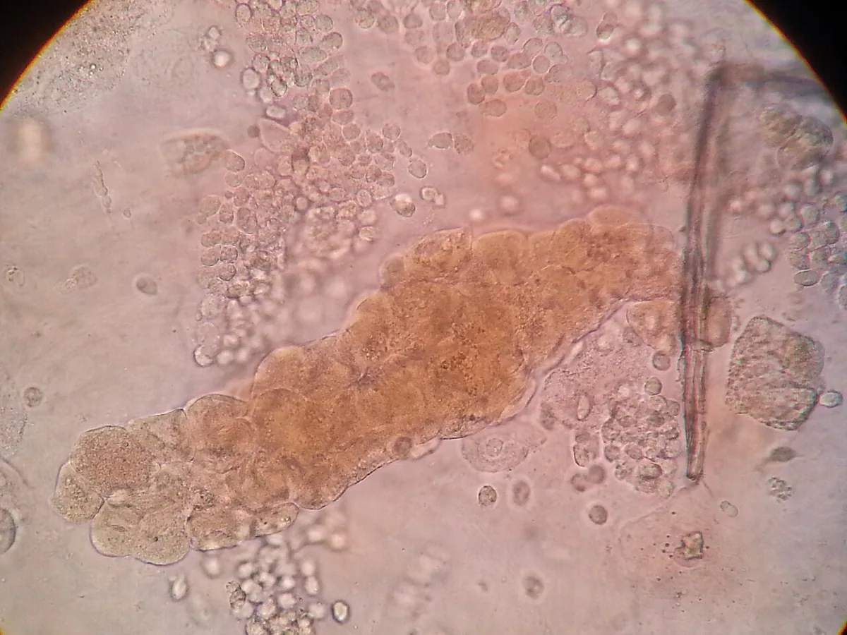
The urine sediment is bland in prerenal AKI. Tubular epithelial cells and numerous, often brown-pigmented granular casts indicate tubular injury; red blood cell casts and dysmorphic red cells indicate glomerulonephritis or vasculitis.
Imaging
- Postvoid residual volume: > 100 mL indicates inadequate bladder emptying.
- Renal ultrasound: hydronephrosis, kidney size (small kidneys suggest chronic kidney disease). Early obstruction or obstruction with hypovolemia may be missed.
- Noncontrast CT to localize an obstruction, magnetic resonance angiography if renal artery stenosis or vascular thrombosis is suspected
Serology and biopsy
If an intrinsic renal cause is suspected: anti-GBM antibodies, ANCA, antinuclear and anti-dsDNA antibodies, antistreptolysin O titer and complement (C3, C4). If the cause remains unclear, a kidney biopsy clarifies it.
Keep learning in the app
Further reading (open access)
Cross-references
More topics: Nephrology
- Nephrotic syndrome
- Chronic kidney disease
- Nephritic syndrome and glomerulonephritis
- Acute interstitial nephritis
- Goodpasture syndrome (anti-GBM disease)
- Membranous nephropathy
- Renal artery stenosis
- Diabetic kidney disease
- Urinary tract infection and cystitis
- Acute pyelonephritis
- Autosomal dominant polycystic kidney disease (ADPKD)
- Hantavirus infection (nephropathia epidemica)
Note: Learning content for medical education – not a treatment recommendation and no substitute for diagnosis or treatment decisions in individual cases. Treatment and management are deliberately not covered on this page.