Autosomal dominant polycystic kidney disease (ADPKD)
Board exam relevance: in 1 of 105 exam reports · rank 181- Synonyms
- polycystic kidney disease, PKD, cystic kidneys, polycystic kidneys
- Specialty
- Internal medicine · Nephrology
- Images
- Ultrasound 1 · Gross specimen 2
- Last updated
- 10/2026 · Dr. Pascal Bafteh
Contents
Images (3)
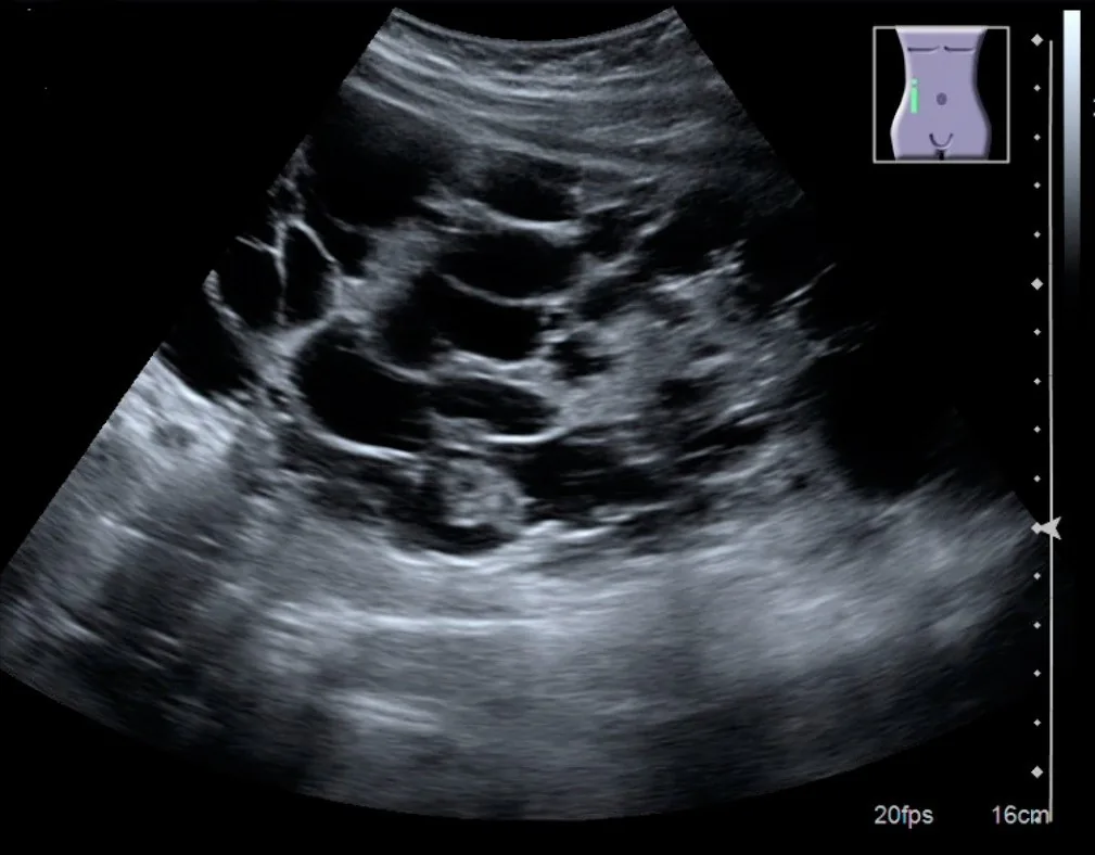 Ultrasound
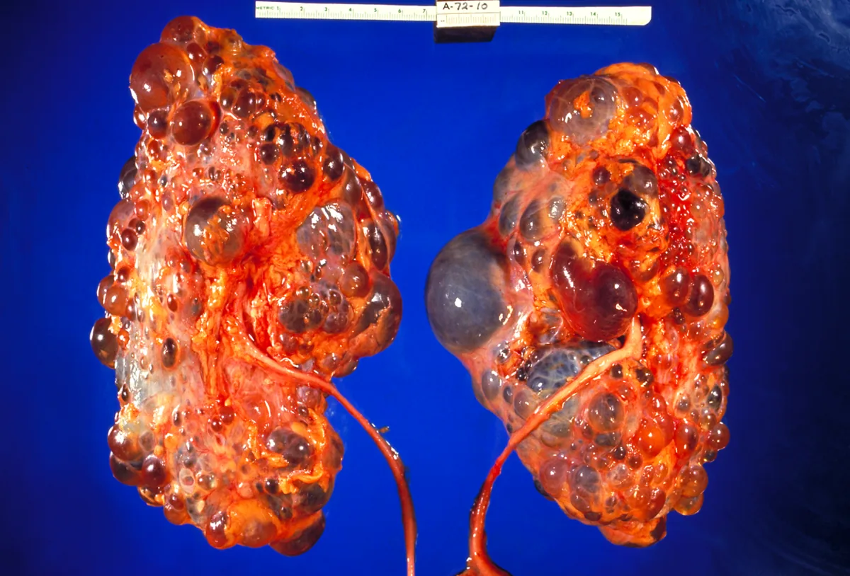 Gross specimen
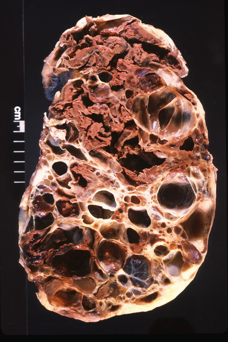 Gross specimen
Definition
Autosomal dominant polycystic kidney disease (ADPKD) is a hereditary disease in which cysts progressively form in both kidneys. The kidneys enlarge, functional tissue is displaced, and kidney failure may develop over time. Other organs are frequently involved.
Classification
Mayo classification
- The Mayo Imaging Classification relates height-adjusted total kidney volume (htTKV, preferably measured by MRI) to age.
- Typical forms (class 1) are graded by the estimated annual volume increase: 1A < 1.5%, 1B 1.5–3%, 1C 3–4.5%, 1D 4.5–6%, 1E > 6%; 1E corresponds to the most rapid progression.
- Atypical forms (class 2A and 2B, e.g. unilateral, segmental or asymmetric) cannot be graded by kidney volume.
- PROPKD score: male sex 1 point, hypertension before age 35 2 points, first urologic event before age 35 2 points, genotype (PKD2 0, nontruncating PKD1 variant 2, truncating PKD1 variant 4 points)
Occurrence & epidemiology
The incidence is about 1 in 1000; ADPKD accounts for about 5% of patients with end-stage kidney disease. Clinical signs are rare before adulthood, but penetrance is essentially complete. The autosomal recessive form (ARPKD) is much rarer at about 1 in 10,000 and usually presents in childhood.
Aetiopathogenesis
Genetics
- Inheritance: autosomal dominant
- Main genes: mostly PKD1, in most remaining cases PKD2; a few familial cases are unrelated to either locus.
- PKD1 variants, especially truncating ones, usually run a more severe course than PKD2 variants.
- Distinction from ARPKD: autosomal recessive (mostly the PKHD1 gene), typically presenting in utero or in infancy
Pathogenesis
PKD1 on chromosome 16 encodes polycystin 1, PKD2 on chromosome 4 encodes polycystin 2. Dysfunction of the primary cilia, with which tubular cells sense urine flow, is thought to promote cell proliferation and cyst formation. Vasopressin increases cell growth and fluid secretion into the cysts via the cAMP pathway. The tubules dilate, separate from the nephron and fill with secreted fluid. By age 60, about 35–45% develop kidney failure.
Clinical features
The disease is asymptomatic for a long time; about half of affected people remain symptom-free throughout life and are never diagnosed. Those who develop symptoms usually do so by the end of their 20s.
- Renal: dull flank, abdominal and back pain due to enlarged kidneys; acute pain from cyst hemorrhage or passage of a stone; hematuria and hypertension each in about 40–50%; subnephrotic proteinuria in about 20%; pyelonephritis, cyst infections and kidney stones; palpably enlarged kidneys in advanced stages
- Liver and abdomen: liver cysts in most patients (liver function usually normal), pancreatic and intestinal cysts, colonic diverticula, inguinal and abdominal wall hernias
- Heart and vessels: valvular abnormalities (mostly mitral valve prolapse, aortic regurgitation) in 25–30% on echocardiography; intracranial aneurysms in about 4% of young and up to 10% of older adults, rupture usually before age 50
Anemia is less common than in other chronic kidney diseases because erythropoietin production is preserved.
Diagnosis
- Ultrasound as the first test: numerous bilateral cysts, enlarged kidneys. With a positive family history, age- and cyst-number-based ultrasound criteria are used; without a family history, no imaging criteria are established.
- CT or MRI: more sensitive than ultrasound, helpful in equivocal cases and for measuring kidney and cyst volume
- Genetic testing if there is no family history, imaging is inconclusive or in younger persons (about under 30 years), in whom imaging is often inconclusive
- Urine and blood: mild proteinuria, microscopic or gross hematuria; pyuria is common even without infection, so infection is confirmed by culture and clinical findings. Creatinine and urea rise slowly; rarely polycythemia.
- Cerebral vessels: magnetic resonance angiography or high-resolution CT in case of symptoms of an aneurysm or a family history of hemorrhagic stroke or aneurysm
Keep learning in the app
Further reading (open access)
Cross-references
More topics: Nephrology
- Acute kidney injury
- Nephrotic syndrome
- Chronic kidney disease
- Nephritic syndrome and glomerulonephritis
- Acute interstitial nephritis
- Goodpasture syndrome (anti-GBM disease)
- Membranous nephropathy
- Renal artery stenosis
- Diabetic kidney disease
- Urinary tract infection and cystitis
- Acute pyelonephritis
- Hantavirus infection (nephropathia epidemica)
Note: Learning content for medical education – not a treatment recommendation and no substitute for diagnosis or treatment decisions in individual cases. Treatment and management are deliberately not covered on this page.