Renal artery stenosis
Board exam relevance: in 3 of 105 exam reports · rank 111- Synonyms
- renovascular hypertension, renal artery occlusion, fibromuscular dysplasia, renal infarction, RAS
- Specialty
- Internal medicine · Nephrology
- Images
- Angiography 1
- Last updated
- 10/2026 · Dr. Pascal Bafteh
Contents
Images (1)
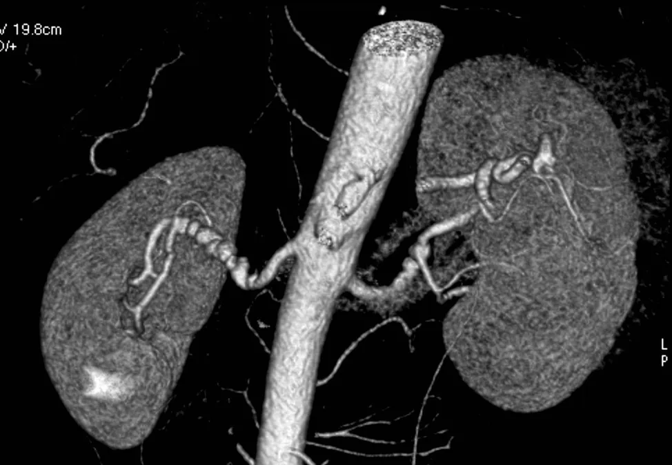 Angiography
Definition
Renal artery stenosis is a narrowing of one or both renal arteries or their branches with reduced blood flow; complete blockage is called renal artery occlusion. Hypoperfusion causes renovascular hypertension, ischemic nephropathy with kidney failure and, with complete occlusion, renal infarction.
Aetiopathogenesis
- Atherosclerosis (about 90% of chronic stenoses): mostly in people older than 45 years, more often men, often bilateral; typically affects the aortic orifice and proximal segment. It often becomes clinically evident after about 10 years of atherosclerosis.
- Fibromuscular dysplasia (almost 10%): irregular thickening of the arterial wall, mostly of the media, typically in the distal main renal artery or intrarenal branches; usually unilateral, mainly in women aged 20 to 50 years
- Rare (< 1%): Takayasu arteritis, Kawasaki disease, neurofibromatosis type 1, aortic dissection or intramural hematoma
- Acute occlusion: usually thromboembolism (atrial fibrillation, after myocardial infarction, endocarditis vegetations, atheroemboli from the aorta), less often thrombosis after trauma or vascular catheterization, aortic dissection; total occlusion for 30–60 minutes causes a wedge-shaped infarction.
Reduced renal perfusion activates the renin-angiotensin-aldosterone system and thus causes renovascular hypertension.
Clinical features
Unilateral stenosis often remains asymptomatic for a long time. Chronic progressive stenosis causes hypertension that begins at an atypical age (before 30 or after 50 years) and may be difficult to control despite several blood pressure-lowering drugs. An abdominal bruit and signs of generalized atherosclerosis may be present; signs of chronic kidney disease develop slowly.
Acute occlusion causes steady, aching flank and abdominal pain, fever, nausea, vomiting, gross hematuria, oliguria or anuria; after 24 hours, signs of acute kidney injury may develop. With an embolic cause, emboli elsewhere may be present ("blue toe" syndrome, livedo reticularis, retinal lesions).
Diagnosis
Suspicion arises with hypertension before age 30 without a family history, severe or difficult-to-control hypertension, unexplained creatinine elevation, and an acute creatinine rise of at least 50% after starting a drug that blocks the renin-angiotensin system.
- Duplex ultrasonography: noninvasive, provides information on kidney function; operator-dependent, time-consuming, limited in obesity
- CT angiography: fast and widely available, requires iodinated contrast
- Magnetic resonance angiography: highly accurate; with reduced GFR, risk of nephrogenic systemic fibrosis from gadolinium
- Catheter angiography or digital subtraction angiography: diagnostic gold standard, invasive, with a risk of atheroembolism and contrast nephropathy
- Renal scintigraphy: more accurate in unilateral than bilateral stenosis, with relevant false-positive and false-negative rates; usually not used as the initial test
- Laboratory tests: creatinine, urea and potassium to assess kidney function; if embolism is suspected, ECG or Holter ECG (atrial fibrillation) and transesophageal echocardiography
Keep learning in the app
Further reading (open access)
Cross-references
More topics: Nephrology
- Acute kidney injury
- Nephrotic syndrome
- Chronic kidney disease
- Nephritic syndrome and glomerulonephritis
- Acute interstitial nephritis
- Goodpasture syndrome (anti-GBM disease)
- Membranous nephropathy
- Diabetic kidney disease
- Urinary tract infection and cystitis
- Acute pyelonephritis
- Autosomal dominant polycystic kidney disease (ADPKD)
- Hantavirus infection (nephropathia epidemica)
Note: Learning content for medical education – not a treatment recommendation and no substitute for diagnosis or treatment decisions in individual cases. Treatment and management are deliberately not covered on this page.