Primary sclerosing cholangitis (PSC)
Board exam relevance: in 6 of 105 exam reports · rank 59- Synonyms
- PSC, sclerosing cholangitis, small-duct PSC, bile duct scarring
- Specialty
- Internal medicine · Liver & biliary tract
- Images
- Ultrasound 1
- Last updated
- 10/2026 · Dr. Pascal Bafteh
Contents
Images (1)
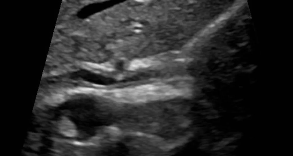 Ultrasound
Definition
PSC is a chronic, progressive cholestatic liver disease. Inflammation and fibrosis lead to strictures and dilatations of the intrahepatic and/or extrahepatic bile ducts. The diagnosis is based on typical bile duct changes on cholangiography after exclusion of other causes of secondary cholangitis.
Classification
- Classic PSC: involvement of large and small bile ducts, frequently with inflammatory bowel disease
- Small-duct PSC: about 5% of patients; only small bile ducts affected, normal cholangiogram, diagnosis by histology only; progression to the classic form is possible
- PSC with features of autoimmune hepatitis (overlap form)
Occurrence & epidemiology
Prevalence is about 10 per 100,000. More than 60% of patients are men; the disease usually manifests between the ages of 30 and 40 but also occurs in children and older people. PSC is found in 2.4–7.5% of patients with ulcerative colitis and in 1.2–3.4% of patients with Crohn's disease.
Aetiopathogenesis
The cause is unknown. Immune-mediated mechanisms are suggested by the close association with inflammatory bowel disease (in 60–80% of PSC patients in Northern Europe and North America) and the frequent detection of autoantibodies such as ANA and atypical pANCA. There is also a genetic component: first-degree relatives have a markedly increased risk of disease.
The accompanying bowel disease often shows a distinct pattern: pancolitis, rectal sparing and backwash ileitis are more common than in ulcerative colitis without PSC.
Clinical features
Up to 50% of patients are symptom-free at diagnosis and are noticed only because of raised liver values. Typical complaints are right upper quadrant pain, pruritus, jaundice, fatigue and exhaustion; episodes of fever point to bacterial cholangitis. On examination, hepatomegaly and splenomegaly are the main findings.
Complications over time:
- bacterial cholangitis (in about 40%) and gallstones or choledocholithiasis (in about 50%)
- dominant strictures of the large bile ducts
- cholangiocarcinoma in 10–20% of patients, and an increased risk of gallbladder carcinoma
- increased risk of colorectal carcinoma with coexisting inflammatory bowel disease
- biliary cirrhosis with portal hypertension
Diagnosis
- Laboratory tests: alkaline phosphatase and GGT usually raised; a normal ALP (in up to 20%) does not exclude PSC, and values often fluctuate. Aminotransferases usually only 2 to 3 times raised, bilirubin usually normal at first.
- Autoantibodies: ANA and pANCA frequently positive but not diagnostic; AMA negative
- IgG4 once at diagnosis to distinguish IgG4-associated cholangitis; however, raised IgG4 levels (above 1.4 g/L) also occur in 10–15% of PSC patients
- MRCP as the most important method of cholangiography: beaded pattern of strictures and upstream dilatations of the bile ducts
- Liver biopsy only if small-duct PSC or overlap with autoimmune hepatitis is suspected; concentric, onion-skin fibrosis around bile ducts is typical
- In known inflammatory bowel disease, persistently raised cholestatic enzymes suggest PSC; however, the bowel disease may also be diagnosed only after the PSC
Keep learning in the app
Further reading (open access)
Cross-references
Note: Learning content for medical education – not a treatment recommendation and no substitute for diagnosis or treatment decisions in individual cases. Treatment and management are deliberately not covered on this page.