Ischemic colitis
Board exam relevance: in 2 of 105 exam reports · rank 142- Synonyms
- colonic ischaemia, colon ischaemia, bowel ischaemia, ischaemic colopathy
- Specialty
- Internal medicine · Gastroenterology
- Images
- CT 1 · Histology 1
- Last updated
- 10/2026 · Dr. Pascal Bafteh
Contents
Images (2)
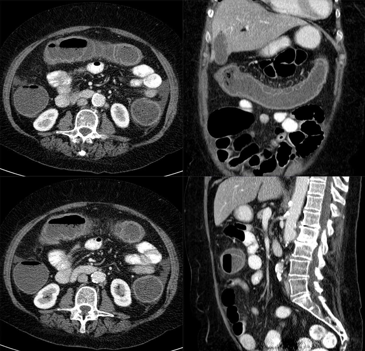 CT
CT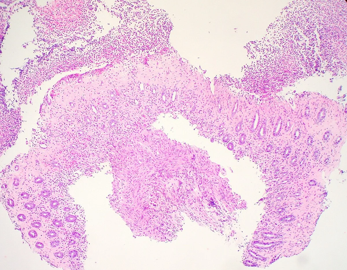 Histology
Definition
Ischemic colitis comprises conditions with insufficient blood supply to the colon due to occlusive or non-occlusive vascular disease. It is usually a transient reduction in blood flow; necrosis is generally limited to the mucosa and submucosa and only occasionally involves the full thickness of the wall. It is the most common form of intestinal ischemia.
Classification
- According to Marston: gangrenous, stricturing and transient forms.
- According to Brandt and Boley (degree of histological damage): reversible colopathy, transient colitis, chronic colitis, stricture, fulminant universal colitis and gangrene.
- Isolated right-sided ischemia: often linked to stenosis or occlusion of the superior mesenteric artery; particularly in sepsis, hypotension, shock or chronic kidney failure.
Occurrence & epidemiology
The age- and sex-adjusted incidence is about 16.3 per 100,000 population and has increased almost fourfold over 34 years. Frequency rises steeply with age – from about 1.1 per 100,000 in people under 40 to about 107 per 100,000 in those over 80. The disease mainly affects people over 60–65 years, women more often than men.
Aetiopathogenesis
- Vascular disease: thought to be mainly small-vessel atherosclerosis; comorbidities such as hypertension, diabetes, kidney disease and coronary artery disease are common.
- Cardiac emboli: about 35 % of patients with segmental, non-gangrenous colitis have a proven cardiac source of embolism, mainly atrial fibrillation.
- Circulatory failure: e.g. myocardial infarction with cardiogenic shock.
- Postoperative: as a complication after abdominal aortic aneurysm repair and after cardiac procedures.
- Triggering substances: including agents that increase urine output, antihypertensive drugs, digoxin, NSAIDs, oral contraceptives, pseudoephedrine, interferon, cocaine.
- Other factors: hereditary coagulation disorders, strenuous physical activity, abdominal fat accumulation, smoking (also in younger people), constipation with raised intraluminal pressure; frequent laxative use may aggravate the condition.
Clinical features
- Cardinal symptoms: abdominal pain, hematochezia and diarrhea. The pain is of acute, cramping onset, often with an urge to defecate, typically in the left lower quadrant; symptoms are milder and of slower onset than in acute mesenteric ischemia.
- Bleeding: bloody stools usually within 24 hours, generally not severe.
- Other symptoms: loss of appetite (eating worsens the pain), abdominal distension, nausea and vomiting; in severe cases a systemic inflammatory response with tachypnea and tachycardia up to sepsis.
- Location: most often the left colon, followed by the distal colon, right colon, transverse colon and pancolon.
- Right-sided ischemia: usually severe abdominal pain with hematochezia and/or diarrhea; bleeding may also be absent.
- Physical findings: mild to severe tenderness over the affected segment; in gangrenous colitis severe tenderness, rebound pain, fever, reduced or absent bowel sounds, shock.
Histology
Common findings are hemorrhage and edema of the mucosa and submucosa, erosions, granulation tissue, gland atrophy, hemosiderin-laden macrophages and an inflammatory infiltrate of the lamina propria. Infarction and "ghost cells" are the most specific but rare findings.
Diagnosis
- Laboratory tests: blood count, electrolytes and metabolic panel, lactate, LDH, D-dimer, CK, amylase, stool culture and Clostridioides difficile toxin; these lack sensitivity and specificity and are of limited help for early diagnosis. In younger patients and recurrences, coagulation disorders are sought.
- CT or colonoscopy: confirm the diagnosis. Typical CT findings are wall thickening, edema, thumbprinting, bowel dilatation and pericolic fluid, sometimes a double-halo sign; pneumatosis and portal venous gas suggest transmural ischemia.
- CT angiography: when acute mesenteric ischemia or isolated right-sided ischemia is suspected.
- Colonoscopy with biopsy: the gold standard, performed early after symptom onset if hemodynamically stable and without peritonitis. Typical findings are edematous, friable mucosa, segmental erythema, petechial hemorrhages and longitudinal ulcers with a segmental, patchy distribution; the "single-stripe" sign (a single longitudinal line of erythema with erosion or ulcer) is highly specific.
- Ultrasound: detects early wall changes and may be an alternative in high-risk patients; wall thickening, however, is non-specific.
- Abdominal X-ray: no diagnostic advantage, but excludes perforation or obstruction; barium enemas have largely been abandoned.
Keep learning in the app
Further reading (open access)
Cross-references
More topics: Gastroenterology
Note: Learning content for medical education – not a treatment recommendation and no substitute for diagnosis or treatment decisions in individual cases. Treatment and management are deliberately not covered on this page.