Helicobacter pylori infection
Board exam relevance: in 2 of 105 exam reports · rank 142- Synonyms
- H. pylori, Helicobacter, HP infection, stomach bug, type B gastritis
- Specialty
- Internal medicine · Gastroenterology
- Images
- Endoscopy 1 · Histology 3
- Last updated
- 10/2026 · Dr. Pascal Bafteh
Contents
Images (4)
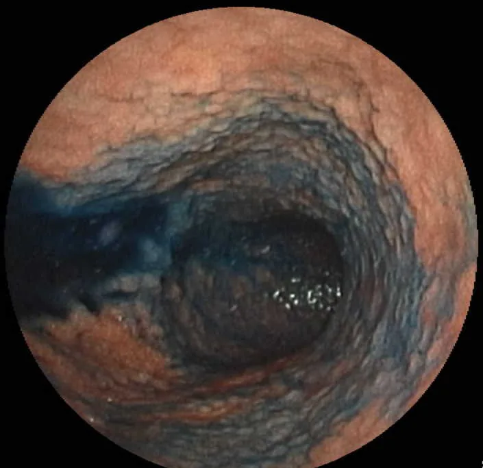 Endoscopy
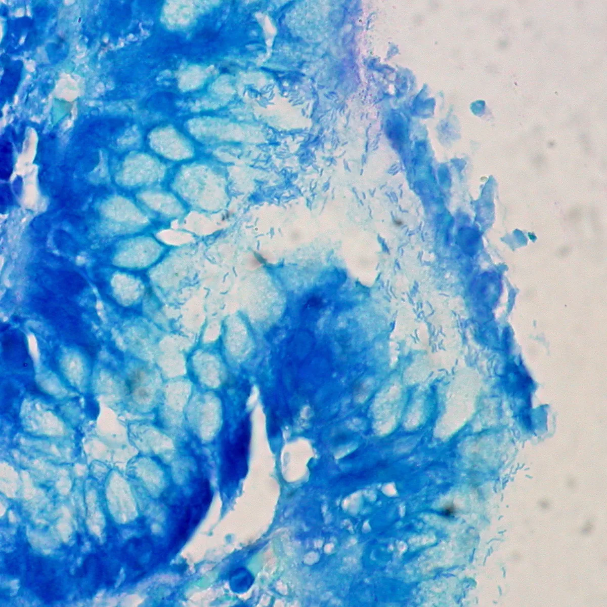 Histology
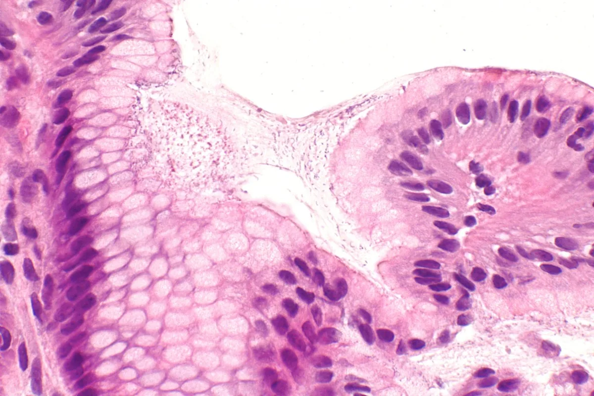 Histology
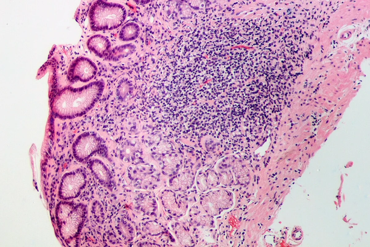 Histology
Definition
Helicobacter pylori is a spiral-shaped, Gram-negative bacterium adapted to the acidic environment of the stomach. Infection causes gastritis, peptic ulcers, gastric adenocarcinoma and low-grade gastric lymphoma. It may be asymptomatic or cause dyspepsia of varying severity. H. pylori is classified as a group 1 carcinogen.
Occurrence & epidemiology
In low- and middle-income countries, H. pylori commonly causes chronic infections, usually acquired in childhood. In North America, prevalence is estimated at 30–40 %; infection is less common in children and increases with age. Infections cluster in families and in residents of custodial institutions.
Aetiopathogenesis
- Transmission: the organizm has been cultured from stool, saliva and dental plaque, suggesting oral–oral or fecal–oral transmission; transmission via inadequately cleaned endoscopes is also possible.
- Ammonia production: enables persistence in the acidic stomach and may erode the mucus barrier; cytotoxins and mucolytic enzymes contribute to mucosal damage.
- Antrum-predominant infection: increased gastrin production (probably via impaired local regulation) with acid hypersecretion; predisposes to prepyloric and duodenal ulcers.
- Body-predominant infection: gastric atrophy with reduced acid production; predisposes to gastric ulcers and gastric adenocarcinoma.
- Ulcer risk: about 10 % of infected people develop a peptic ulcer, compared with 1 % of uninfected people.
- Malignancy: association with intestinal-type adenocarcinoma of the gastric body and antrum (not cardia cancer) and with gastric lymphomas including MALT lymphoma.
Clinical features
- No symptoms: many infected people have no noticeable clinical effects.
- Dyspepsia of varying severity.
- Sequelae: chronic gastritis, peptic ulcers with their complications, gastric adenocarcinoma, gastric lymphomas.
Histology
Superficial gastritis in H. pylori infection shows an infiltrate of lymphocytes and plasma cells mixed with neutrophils. Over time, atrophy and mucous gland and intestinal metaplasia may develop. Immunohistochemistry can detect even very small numbers of organizms.
Diagnosis
- Urea breath test and stool antigen test: the preferred tests for initial diagnosis. In the breath test, ¹³C- or ¹⁴C-labelled urea is ingested; the released labelled CO₂ is measured in breath samples taken 20–30 minutes later. Both tests have high sensitivity and specificity.
- Confounders: false-negative results after recent intake of antibacterial drugs or during intake of acid-suppressing drugs; histamine H2 receptor antagonists do not affect the breath test.
- Serology: with declining prevalence often falsely positive and too unreliable in most regions; qualitative assays remain positive for up to 3 years after successful clearance.
- Invasive tests: biopsies when endoscopy is performed for other reasons; the rapid urease test is the preferred method on tissue, and histology complements it when the result is negative but suspicion is high. Culture is of limited use because of the organizm's fastidious growth requirements.
Keep learning in the app
Further reading (open access)
Cross-references
More topics: Gastroenterology
Note: Learning content for medical education – not a treatment recommendation and no substitute for diagnosis or treatment decisions in individual cases. Treatment and management are deliberately not covered on this page.