Goiter and thyroid nodules
Board exam relevance: in 3 of 105 exam reports · rank 111- Synonyms
- thyroid enlargement, thyroid lump, nodular goitre, multinodular goiter, goiter, endemic goitre
- Specialty
- Internal medicine · Endocrinology & diabetes
- Images
- Clinical 2 · Histology 1
- Last updated
- 10/2026 · Dr. Pascal Bafteh
Contents
Images (3)
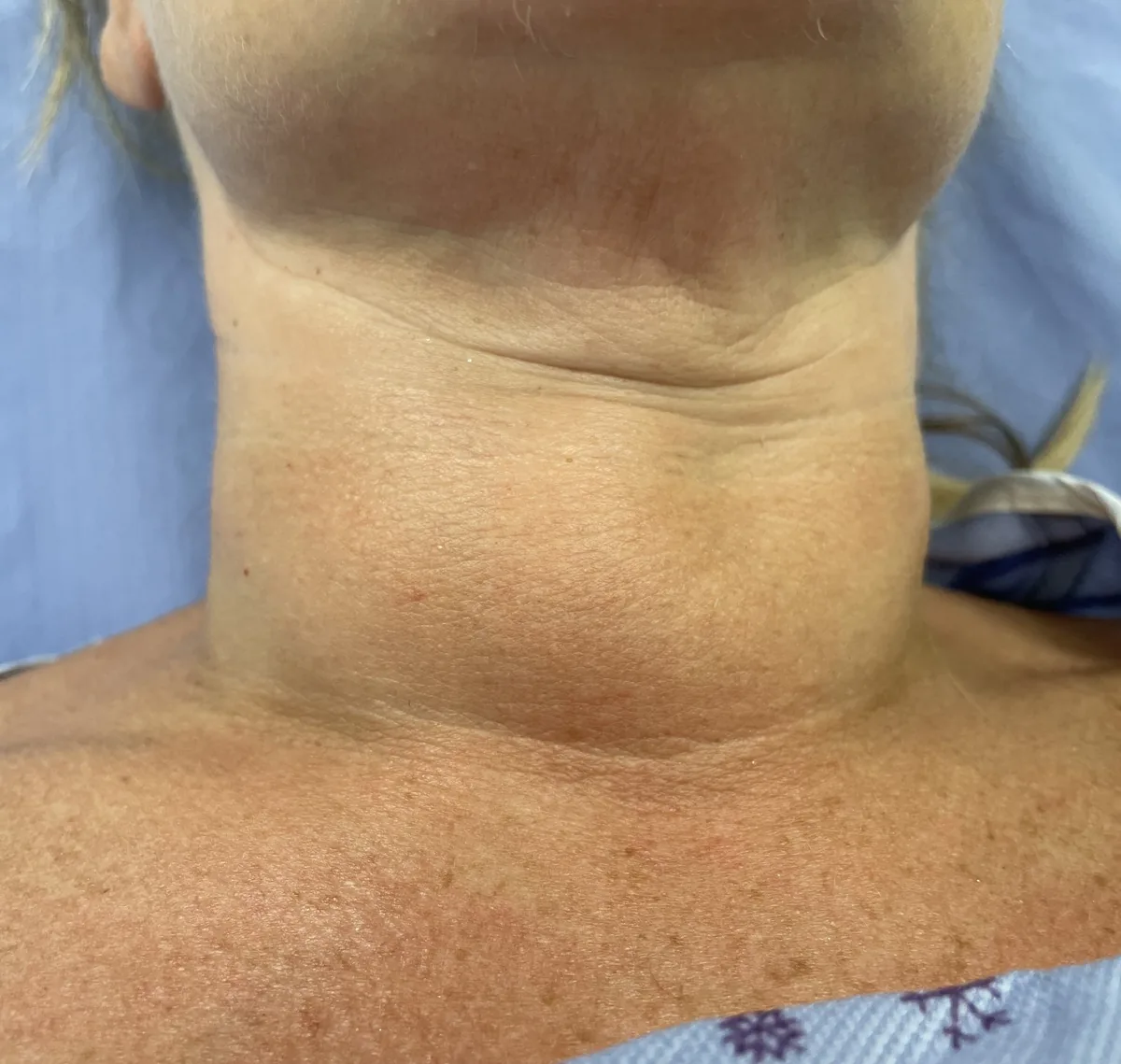
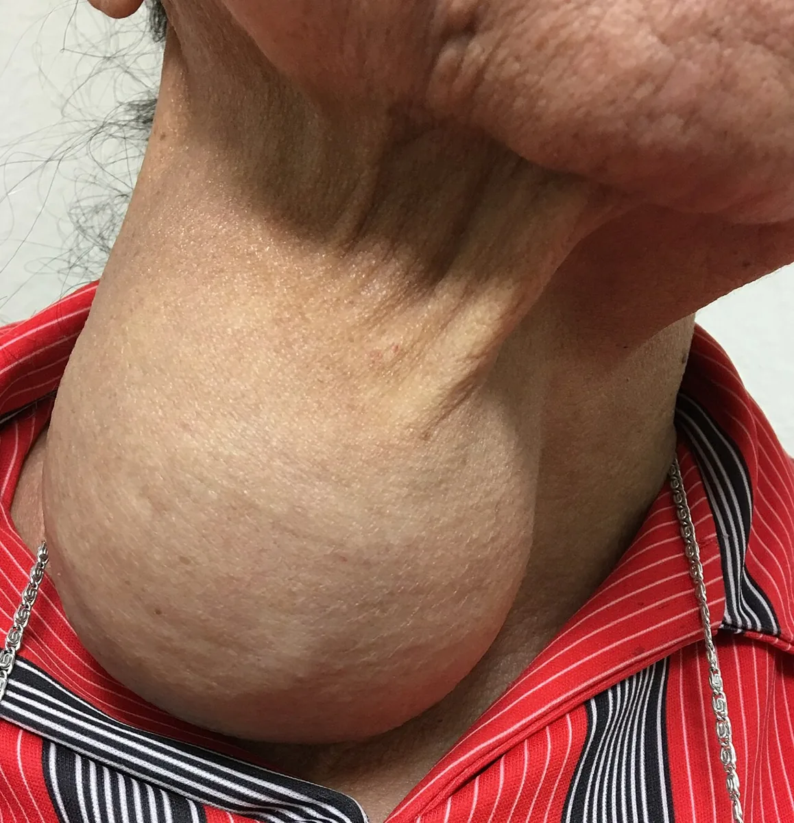
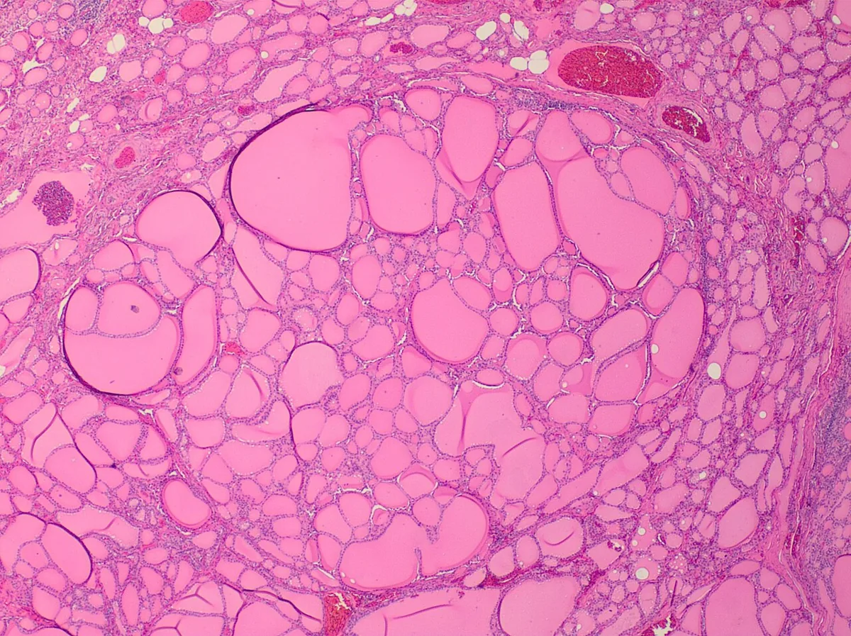 Histology
Definition
A goiter is any enlargement of the thyroid gland. It may be uniform (diffuse goiter) or nodular (nodular goiter). Simple (euthyroid) goiter is a non-cancerous enlargement without hyperthyroidism, hypothyroidism or inflammation; it is the most common type of thyroid enlargement.
A thyroid nodule is a discrete lesion within the thyroid that is palpably or radiologically distinct from the surrounding tissue. Most nodules are benign; the key question is distinguishing them from thyroid cancer.
Classification
- By morphology: diffuse goiter; uninodular or multinodular goiter; cystic components.
- By function: euthyroid (simple goiter), hyperthyroid (e.g. hyperfunctioning adenoma) or hypothyroid (e.g. Hashimoto thyroiditis).
- By scintigraphy: nodules with increased radionuclide uptake ("hot" nodules) are seldom malignant.
- TI-RADS: malignancy risk classification based on ultrasound features – size, echogenicity, composition, shape, margins and echogenic foci.
- Bethesda system (cytology): category II benign, III and IV indeterminate, V suspicious for malignancy, VI malignant.
Occurrence & epidemiology
Iodine deficiency is the most common cause of goiter worldwide (endemic goiter). Simple goiter is often first noticed at puberty, during pregnancy and at menopause.
The frequency of thyroid nodules depends on the method of assessment: in middle-aged and older people about 5 % are palpable, while ultrasound and autopsy studies find nodules in about 50 % of older adults. Many nodules are discovered incidentally on neck imaging done for other reasons.
Aetiopathogenesis
- Iodine deficiency: a slight compensatory rise in TSH keeps thyroid function within the normal range but stimulates thyroid growth. Recurrent cycles of stimulation and involution lead to nodule formation.
- Defects of hormone synthesis: intrinsic defects of thyroid hormone production.
- Goitrogenic foods: e.g. in cassava, cabbage, broccoli and cauliflower, mainly in iodine-deficient regions.
- Certain substances: such as iodine-containing compounds that inhibit hormone synthesis.
- Unexplained: in iodine-sufficient regions the cause of most simple goiters remains unknown.
Benign causes of nodules: hyperplastic colloid nodules, cysts, thyroiditis, adenomas. Malignant cause: thyroid cancer.
Clinical features
Symptoms
Most patients are asymptomatic; the thyroid is visibly or palpably enlarged and not tender. Initially the goiter is usually soft, symmetric and smooth; later nodules and cysts develop. Large goiters may cause dysphagia, hoarseness or a sensation of fullness in the throat (globus).
Nodules are usually asymptomatic as well. Clues from the history:
- Pain suggests thyroiditis or hemorrhage into a cyst.
- Palpitations, heat intolerance, weight loss and tremor suggest a hyperfunctioning adenoma or thyroiditis.
- Cold intolerance, weight gain and fatigue suggest Hashimoto thyroiditis.
- Compressive symptoms (difficulty swallowing or breathing, hoarseness) increase the likelihood of a malignant nodule.
Features suggesting malignancy
- Risk factors for cancer: radiation exposure of the thyroid, especially in infancy or childhood; age over 55 years; female sex; family history of thyroid cancer or multiple endocrine neoplasia type 2; solitary nodule; increase in size, particularly rapid growth; higher TSH.
- Findings suggestive of malignancy: stony-hard consistency, fixation to surrounding structures, cervical lymphadenopathy and hoarseness due to recurrent laryngeal nerve palsy.
Diagnosis
- TSH: the first laboratory step. Thyroid function is usually normal in simple goiter. In endemic goiter TSH may be slightly elevated, T4 low-normal or slightly low and T3 normal or slightly elevated.
- Thyroid antibodies: to exclude Hashimoto thyroiditis.
- Scintigraphy: with a low TSH, to detect hyperfunctioning ("hot") nodules.
- Ultrasound: determines the size and number of nodules. Suspicious features are hypoechogenicity, marked internal vascularity, irregular margins, a nodule taller than it is wide, irregular macrocalcifications, fine stippled microcalcifications (papillary carcinoma) and dense, homogeneous irregular calcification (medullary carcinoma).
- Ultrasound-guided fine-needle aspiration with cytology: the standard method for distinguishing benign from malignant nodules; reported using the Bethesda system. For cytologically indeterminate results, molecular analysis of the aspirate or measurement of calcitonin (medullary carcinoma) can help.
Keep learning in the app
Further reading (open access)
Cross-references
Note: Learning content for medical education – not a treatment recommendation and no substitute for diagnosis or treatment decisions in individual cases. Treatment and management are deliberately not covered on this page.