Gastro-esophageal reflux disease (GERD)
Board exam relevance: in 2 of 105 exam reports · rank 142- Synonyms
- heartburn, acid reflux, reflux oesophagitis, GERD, GORD, acid indigestion
- Specialty
- Internal medicine · Gastroenterology
- Images
- Endoscopy 1 · X-ray 1 · Histology 1
- Last updated
- 10/2026 · Dr. Pascal Bafteh
Contents
Images (3)
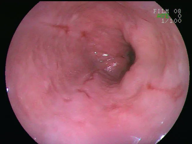 Endoscopy
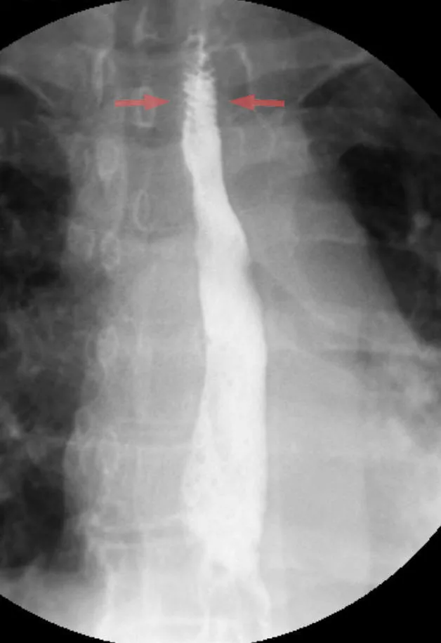 X-ray
X-ray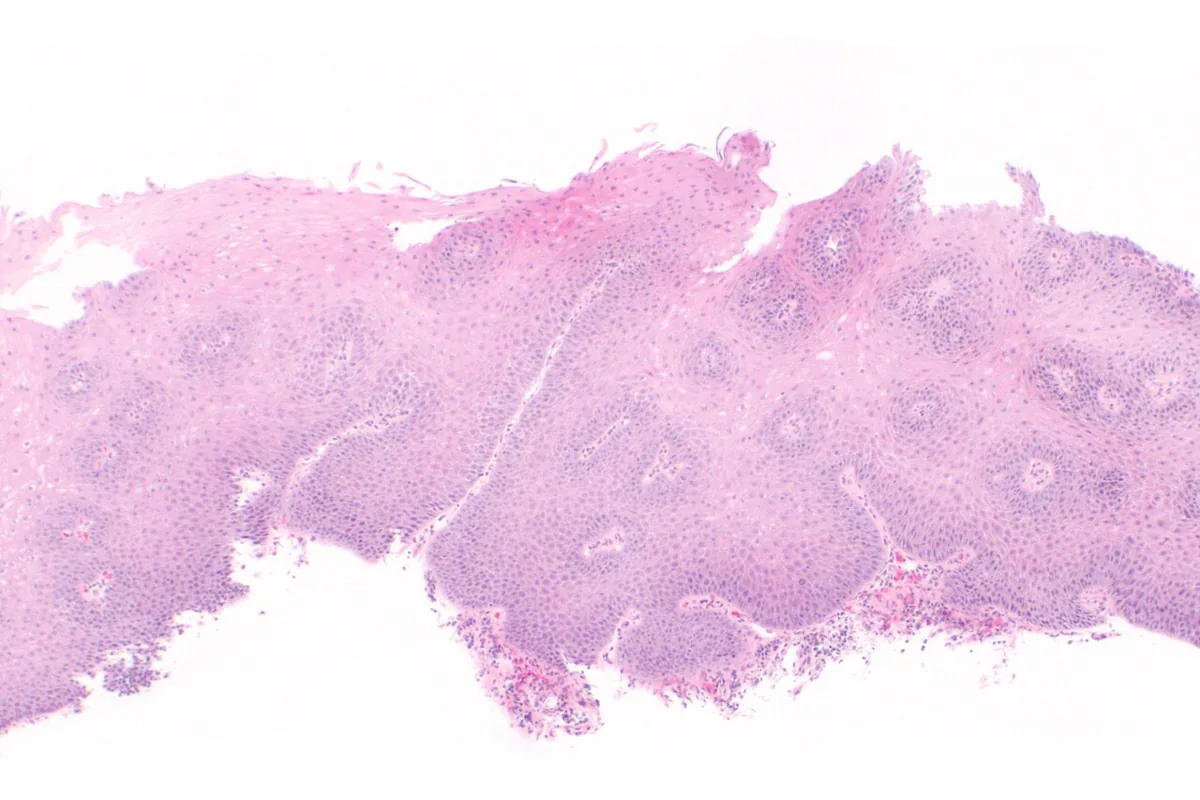 Histology
Definition
In gastro-esophageal reflux disease (GERD), an incompetent lower esophageal sphincter allows gastric contents to flow back into the esophagus, causing burning pain. Prolonged reflux can lead to esophagitis and stricture, and rarely to metaplasia (Barrett's esophagus) or cancer.
Gastro-esophageal reflux is also common in infants, but there it is not always pathological and not automatically GERD.
Classification
Endoscopic grading of reflux esophagitis according to the Los Angeles classification:
- Grade A: one or more mucosal breaks ≤ 5 mm that do not extend across the tops of two mucosal folds.
- Grade B: one or more mucosal breaks > 5 mm that do not extend across the tops of two mucosal folds.
- Grade C: mucosal breaks that extend across at least two folds but involve less than 75 % of the circumference.
- Grade D: mucosal breaks involving ≥ 75 % of the circumference.
According to the Lyon Consensus, grades B, C and D are objective evidence of GERD; with normal endoscopy or only grade A, advanced pH testing is required.
Occurrence & epidemiology
GERD is common and affects about 13 % of the population worldwide. Prevalence varies by region and increases with age. Up to about 94 % of people with GERD have some degree of hiatus hernia.
Aetiopathogenesis
- Incompetence of the lower esophageal sphincter: through a generalised loss of sphincter tone or through recurrent, inappropriate transient relaxations unrelated to swallowing. Transient relaxations are triggered by gastric distension or subthreshold pharyngeal stimulation.
- Protective factors at the junction: the angle between esophagus and cardia, the action of the diaphragm, gravity (upright position) and age.
- Contributing factors: obesity, smoking, fatty foods, caffeinated or carbonated drinks, alcohol; genetic predisposition.
- Lowering of sphincter pressure: by certain drugs, e.g. anticholinergics, histamine H1 receptor antagonists, tricyclic antidepressants, calcium channel blockers, progesterone, nitrates.
- Development of esophagitis: depends on the caustic nature of the refluxate, the ability of the esophagus to clear it, the volume of gastric contents and local mucosal protective mechanisms.
Clinical features
- Cardinal symptom: heartburn, with or without regurgitation of gastric contents into the mouth.
- With chronic aspiration: cough, hoarseness, wheezing.
- Esophagitis: odynophagia and bleeding, usually occult, occasionally massive; over time iron deficiency anemia.
- Peptic stricture: gradually progressive dysphagia for solid food.
- Peptic esophageal ulcer: pain similar to gastric or duodenal ulcer, usually localised to the xiphoid or high retrosternal region; heals slowly, tends to recur and often leaves a stricture.
- Complications: esophagitis, esophageal ulcer, stricture, Barrett's esophagus and esophageal adenocarcinoma.
Diagnosis
- History: a detailed history points to the diagnosis; typical symptoms are heartburn and regurgitation.
- Further assessment: when symptoms do not improve, are long-standing, suggest complications, or overlap with other disorders.
- Endoscopy with cytological washings and/or biopsy of abnormal areas: the preferred test; only biopsy reliably detects the columnar mucosa of Barrett's esophagus.
- Advanced pH testing: when endoscopy is normal and typical symptoms persist; performed off acid-suppressing drugs.
- Barium swallow: readily shows ulcers and peptic strictures but is of little help in mild to moderate reflux disease.
- Esophageal manometry: assesses esophageal peristalsis.
Keep learning in the app
Further reading (open access)
Cross-references
More topics: Gastroenterology
Note: Learning content for medical education – not a treatment recommendation and no substitute for diagnosis or treatment decisions in individual cases. Treatment and management are deliberately not covered on this page.