Gastric cancer
Board exam relevance: in 2 of 105 exam reports · rank 142- Synonyms
- stomach cancer, gastric carcinoma, gastric adenocarcinoma, signet ring cell carcinoma, linitis plastica
- Specialty
- Internal medicine · Gastroenterology
- Images
- Endoscopy 1 · X-ray 1 · Histology 1 · Gross specimen 1
- Last updated
- 10/2026 · Dr. Pascal Bafteh
Contents
Images (4)
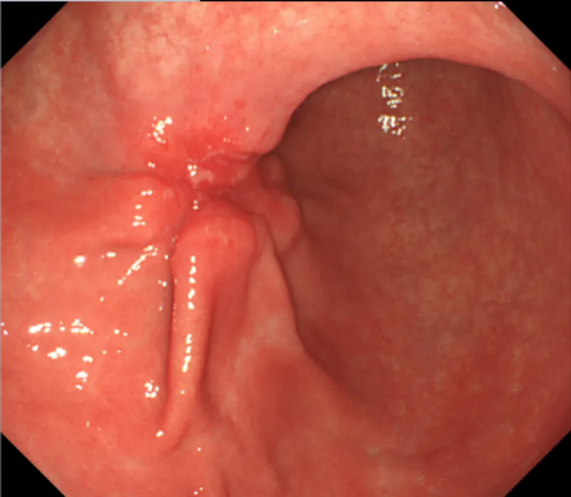 Endoscopy
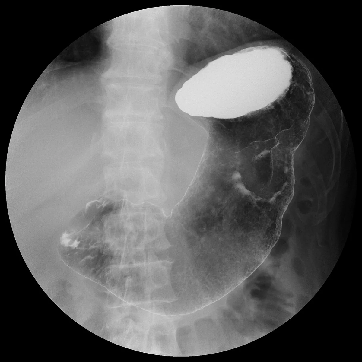 X-ray
X-ray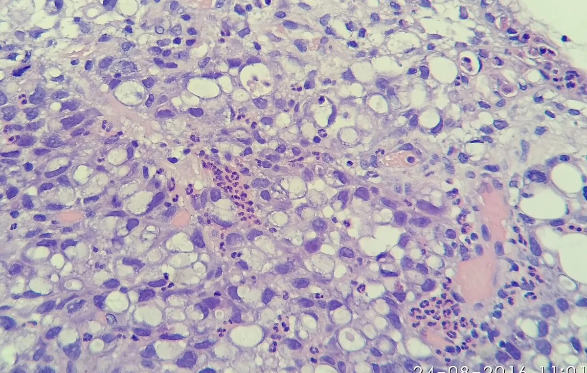 Histology
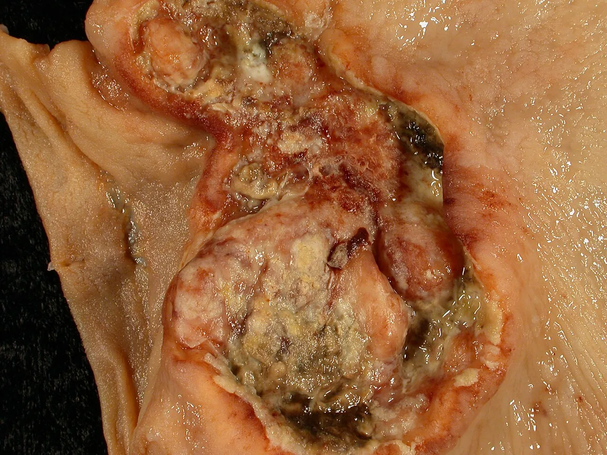 Gross specimen
Definition
Gastric cancer is a malignant tumor of the stomach. Adenocarcinoma accounts for about 95 % of malignant gastric tumors; less common are localised gastric lymphomas, gastrointestinal stromal tumors and leiomyosarcomas.
Classification
Macroscopic classification of adenocarcinoma:
- Protruding: polypoid or fungating tumor.
- Penetrating: ulcerated tumor.
- Superficial spreading: spread along the mucosa or superficial infiltration of the gastric wall.
- Linitis plastica: diffuse infiltration of the gastric wall with a fibrous reaction and a rigid "leather bottle" stomach.
- Miscellaneous: features of two or more types; the largest group.
Occurrence & epidemiology
In Germany, about 9,040 men and 5,580 women were diagnosed with gastric cancer in 2023; the median age at diagnosis is 74 years in women and 71 in men. Incidence and death rates have been falling for decades, most markedly for tumors of the gastric outlet. In men, tumors are located at the cardia about twice as often as in women. In almost 40 % of cases with documented stage, distant metastases are already present at diagnosis.
Worldwide, gastric cancer is the fifth most common cancer, with large regional differences: very high incidence in East Asia and Eastern Europe, lowest in Africa. Frequency increases with age.
Aetiopathogenesis
- Helicobacter pylori: the most important risk factor, especially with extensive intestinal metaplasia.
- Autoimmune atrophic gastritis.
- Epstein–Barr virus: about 5–10 % of gastric cancers.
- Smoking and alcohol.
- Food: salt-preserved foods, high salt intake and processed meat products; for processed meat, the WHO International Agency for Research on Cancer reports a positive association.
- Gastric polyps: adenomatous polyps, especially multiple ones, those over 2 cm or with villous histology, carry the highest risk of malignant change.
- Reflux disease: evidence of an increased risk of tumors at the junction with the esophagus.
- Other factors: postoperative stomach, low socioeconomic status.
- Familial and genetic factors: first-degree relatives have a two- to threefold higher risk. Hereditary diffuse gastric cancer is due to mutations in the CDH1 gene (autosomal dominant, high penetrance; onset at an average age of 38, lifetime risk about 80 %). Other syndromes include familial adenomatous polyposis, Lynch syndrome, juvenile polyposis and Peutz–Jeghers syndrome.
Clinical features
- Early symptoms: non-specific, often dyspepsia resembling peptic ulcer; easily misinterpreted.
- Later: early satiety with obstruction of the pylorus or a non-distensible stomach (linitis plastica), dysphagia with cardia cancer, loss of weight and strength.
- Bleeding: massive hematemesis or melaena is uncommon; anemia from occult blood loss is more frequent.
- Presentation with metastases: e.g. jaundice, ascites or fractures.
- Late findings: epigastric mass, umbilical, left supraclavicular or left axillary lymph nodes, hepatomegaly, ovarian or rectal mass; metastases to lung, central nervous system and bone.
Diagnosis
- Endoscopy with multiple biopsies and brush cytology: when gastric cancer is suspected; a biopsy limited to the mucosa may miss tumor tissue in the submucosa.
- Double-contrast barium study: may show lesions but rarely replaces endoscopy.
- CT of the chest and abdomen: staging once cancer is confirmed.
- Endoscopic ultrasound: if CT shows no metastases, to determine depth of invasion and regional lymph node involvement.
- Laboratory tests: full blood count, electrolytes and liver function tests (anemia, general condition, liver metastases); CEA measurement.
Keep learning in the app
Further reading (open access)
Cross-references
More topics: Gastroenterology
Note: Learning content for medical education – not a treatment recommendation and no substitute for diagnosis or treatment decisions in individual cases. Treatment and management are deliberately not covered on this page.