Focal segmental glomerulosclerosis (FSGS)
- Synonyms
- FSGS, collapsing glomerulopathy, HIV-associated nephropathy
- Specialty
- Internal medicine · Nephrology
- Images
- Histology 2
- Last updated
- 10/2026 · Dr. Pascal Bafteh
Contents
Images (2)
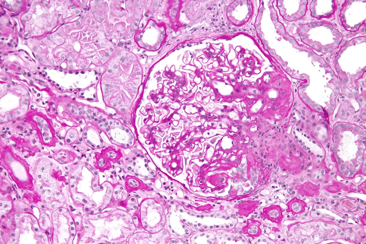 Histology
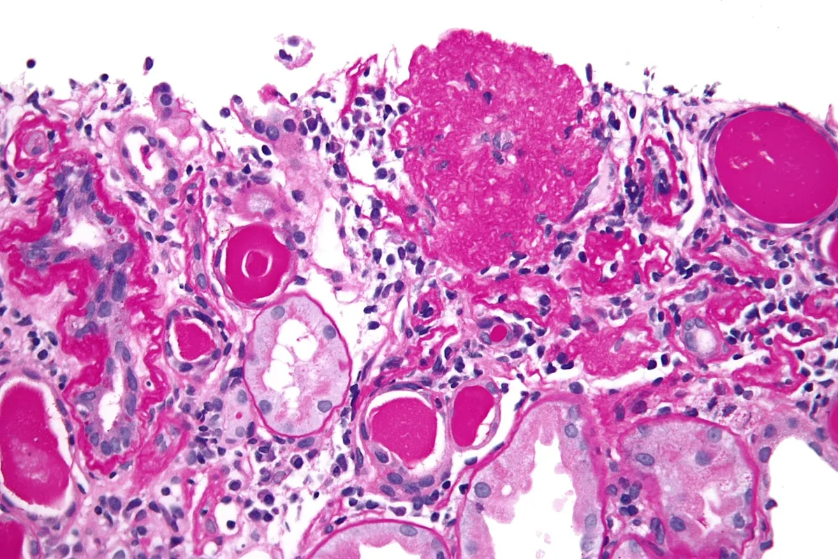 Histology
Definition
Focal segmental glomerulosclerosis (FSGS) is a histologic pattern of injury: only some glomeruli are affected (focal), and within them only part of the capillary tuft (segmental), which becomes scarred and hyalinized. Clinically it usually causes heavy, often nephrotic-range proteinuria.
Occurrence & epidemiology
In the United States, FSGS is the most common cause of idiopathic nephrotic syndrome in adults; Black men are particularly often affected.
Aetiopathogenesis
- Primary (idiopathic): podocyte injury, presumably due to circulating factors
- Familial/genetic: hereditary defects of slit diaphragm proteins
- Secondary adaptive: overload of remaining nephrons with reduced nephron mass (reflux nephropathy, renal dysgenesis, oligomeganephronia) or obesity
- Secondary due to viruses and substances: HIV infection (HIV-associated nephropathy), heroin, certain drugs; also renal atheroembolic disease
The filtration barrier is defective for size and charge; proteinuria is therefore usually nonselective and includes high-molecular-weight proteins as well as albumin.
Clinical features
Typical findings are heavy proteinuria, hypertension, impaired kidney function and edema, alone or in combination. Sometimes there is only asymptomatic proteinuria below the nephrotic range; microscopic hematuria occurs occasionally. The collapsing variant is particularly severe and progresses rapidly to kidney failure.
Histology
- Light microscopy shows focal, segmental sclerosis and hyalinosis of individual glomeruli alongside normal glomeruli. If no affected glomerulus is sampled, the biopsy may be falsely negative.
- Immunofluorescence: coarse granular to nodular deposits of IgM and C3 in sclerotic areas
- Electron microscopy: diffuse effacement of podocyte foot processes in primary FSGS, more patchy in secondary forms
- Variants include the tip lesion (sclerosis at the tubular pole) and the collapsing variant (wrinkled, collapsed capillary loops; typical in HIV or intravenous drug use)
Diagnosis
In unexplained proteinuria, nephrotic syndrome or kidney dysfunction: urinalysis and sediment, creatinine and urea, 24-hour proteinuria or protein-to-creatinine ratio, HIV serology and a history of drug and substance use. The diagnosis is confirmed by kidney biopsy with immunofluorescence and electron microscopy.
Keep learning in the app
Further reading (open access)
Cross-references
More topics: Nephrology
- Acute kidney injury
- Nephrotic syndrome
- Chronic kidney disease
- Nephritic syndrome and glomerulonephritis
- Acute interstitial nephritis
- Goodpasture syndrome (anti-GBM disease)
- Membranous nephropathy
- Renal artery stenosis
- Diabetic kidney disease
- Urinary tract infection and cystitis
- Acute pyelonephritis
- Autosomal dominant polycystic kidney disease (ADPKD)
Note: Learning content for medical education – not a treatment recommendation and no substitute for diagnosis or treatment decisions in individual cases. Treatment and management are deliberately not covered on this page.