Diabetic polyneuropathy
Board exam relevance: in 1 of 105 exam reports · rank 181- Synonyms
- diabetic neuropathy, DSPN, peripheral neuropathy, nerve damage from diabetes, numb feet, burning feet
- Specialty
- Internal medicine · Endocrinology & diabetes
- Images
- Clinical 2
- Last updated
- 10/2026 · Dr. Pascal Bafteh
Contents
Images (2)
Definition
Diabetic polyneuropathy comprises nerve damage caused by diabetes mellitus. The most common form is distal symmetric sensorimotor polyneuropathy (DSPN), which begins in a stocking-and-glove distribution in the feet and hands. It is a major precursor of diabetic foot syndrome.
Classification
- Symmetric polyneuropathy with small-fiber and large-fiber variants
- Autonomic neuropathy
- Radiculopathies: mostly the proximal lumbar roots L2–L4 with pain, weakness and atrophy of the legs (diabetic amyotrophy) or the thoracic roots T4–T12 with abdominal pain
- Cranial neuropathies: of the oculomotor nerve with diplopia, ptosis and anisocoria; less often the trochlear or abducens nerve
- Mononeuropathies: e.g. median or peroneal nerve (foot drop), also as mononeuritis multiplex; in addition a tendency to entrapment syndromes such as carpal tunnel syndrome
Occurrence & epidemiology
Reported frequencies vary with diagnostic criteria and examination methods. According to the Robert Koch Institute, estimates from routine data range from 14 to 28 %, and from 8 to 51 % in studies outside Germany. In the North Rhine-Westphalia DMP (structured care program) in 2020, neuropathy was documented in 28.6 % of people with type 2 diabetes, and in around 40 % of those older than 75 years.
Aetiopathogenesis
Nerve damage results from ischemia due to microangiopathy of the vasa nervorum, from direct effects of hyperglycemia on neurons and from intracellular metabolic changes, for instance via the polyol pathway and the formation of advanced glycation end products. The longest nerve fibers are affected first, which is why DSPN starts at the toes.
Clinical features
Sensorimotor polyneuropathy
- Positive symptoms: tingling and other paresthesias, dysesthesias, pain
- Negative symptoms: painless loss of touch, vibration, position and temperature sense; reduced or absent ankle reflexes
- Small-fiber neuropathy: pain, numbness and loss of temperature sensation with preserved vibration and position sense; tendency to foot ulcers and neuropathic joint destruction
- Large-fiber neuropathy: muscle weakness, loss of vibration and position sense, absent deep tendon reflexes
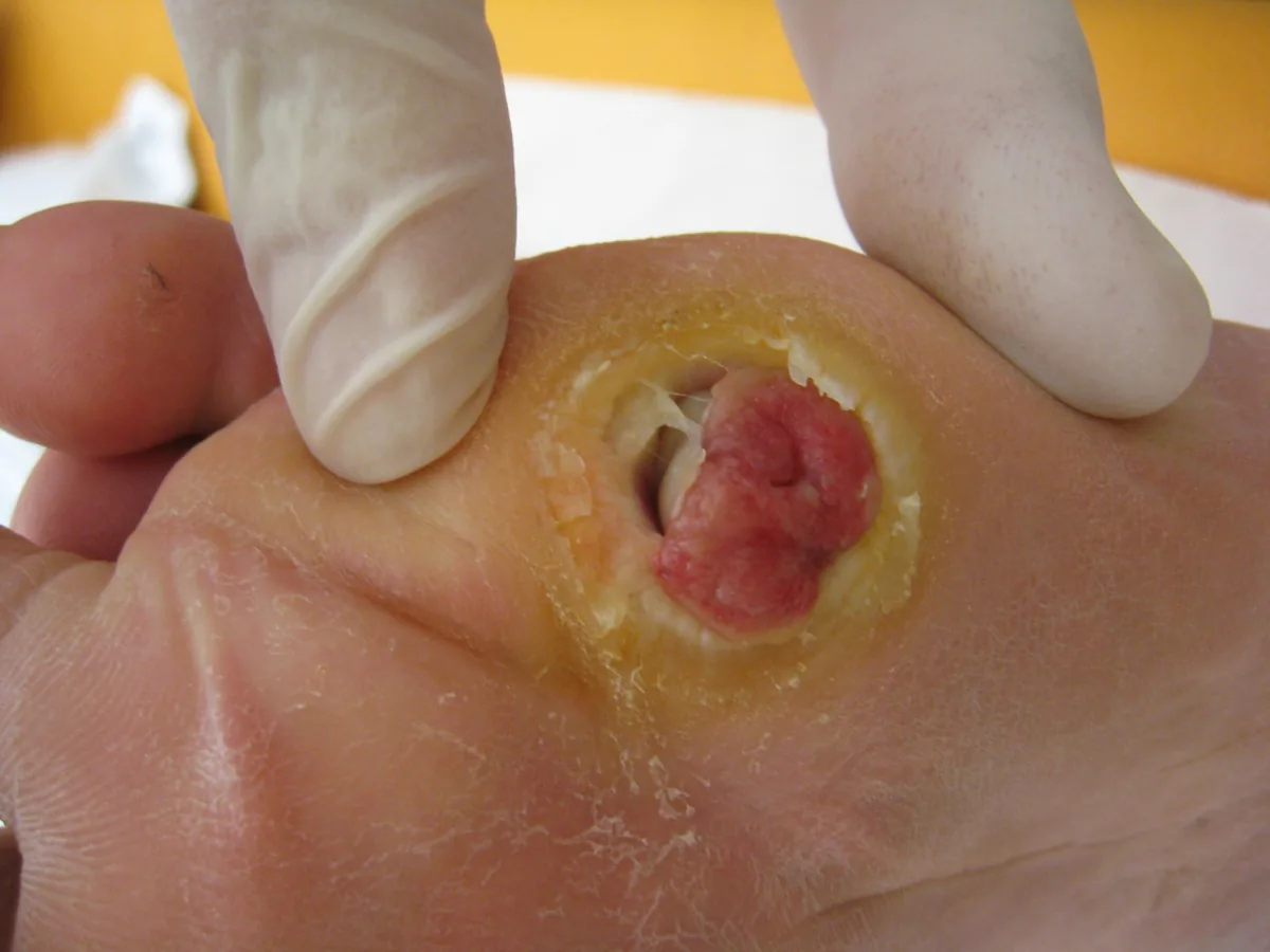
- Consequences: injuries from ill-fitting shoes or abnormal weight bearing go unnoticed; ulcers, infections and fractures develop, up to neuropathic osteoarthropathy (Charcot foot).
Autonomic neuropathy
- Cardiovascular — triad: resting tachycardia + orthostatic hypotension + 'silent ischemia'.
- Gastrointestinal: gastroparesis + diarrhea/constipation.
- Urogenital: bladder-emptying disorder + ED.
- Sudomotor: plantar anhidrosis + compensatory hyperhidrosis.
Diagnosis
The diagnosis of DSPN is based on symptoms, sensory deficits and reduced ankle reflexes. Testing is done on both sides, with the eyes closed and not over calluses, scars or wounds.
- Vibration sense: graduated Rydel-Seiffer tuning fork on the dorsum of the interphalangeal joint of the great toe, and at the medial malleolus if no sensation is felt. The lower limit of normal is age-dependent, at the interphalangeal joint of the great toe e.g. 5/8 up to 39 years and 3.5/8 from 75 years.
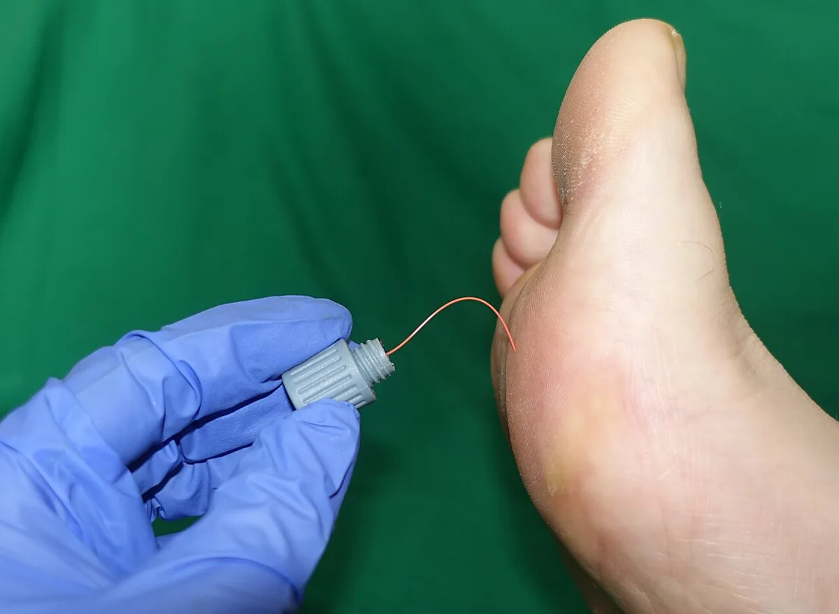
- Pressure and touch sense: 10 g monofilament at a minimum of five plantar sites per foot; abnormal if 1 of 5 or 2 of 10 sites are not detected. Loss of this sensation identifies feet at the highest risk of ulceration.
- Pain sensation: sharp-blunt discrimination on the dorsum of the foot; impaired from 3 of 10 incorrect answers.
- Temperature sense: cold-warm discrimination on the dorsum of the foot; reduced if at least 2 of 3 trials per foot are answered incorrectly.
- Nerve conduction studies and electromyography for atypical presentations and to exclude other causes, such as non-diabetic radiculopathy or carpal tunnel syndrome.
Keep learning in the app
Further reading (open access)
Cross-references
Note: Learning content for medical education – not a treatment recommendation and no substitute for diagnosis or treatment decisions in individual cases. Treatment and management are deliberately not covered on this page.