Autoimmune pancreatitis
Board exam relevance: in 1 of 105 exam reports · rank 181- Synonyms
- AIP, IgG4-related pancreatitis, IgG4-related disease, lymphoplasmacytic sclerosing pancreatitis, idiopathic duct-centric pancreatitis
- Specialty
- Internal medicine · Gastroenterology
- Images
- CT 1 · MRI 1
- Last updated
- 10/2026 · Dr. Pascal Bafteh
Contents
Images (2)
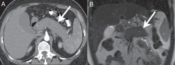 CT
CT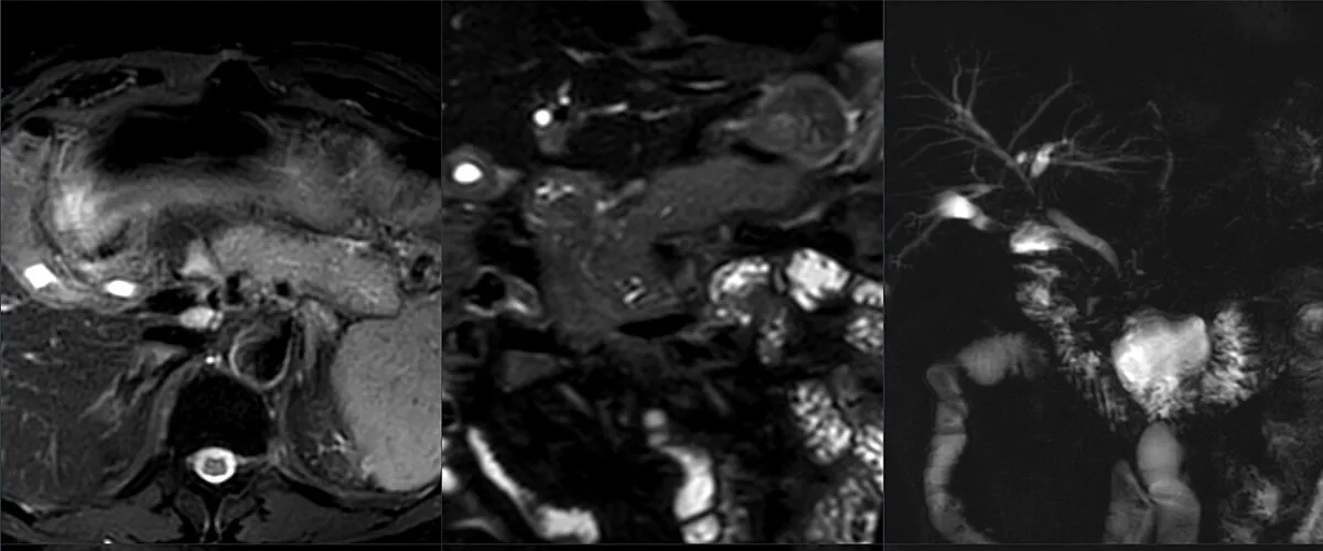 MRI
MRIDefinition
Autoimmune pancreatitis (AIP) is a rare, specific form of chronic benign pancreatic disease. Typical features are obstructive jaundice with or without a pancreatic mass and, histologically, a characteristic lymphoplasmacytic infiltrate with fibrosis. The enlarged or focally altered pancreas can mimic pancreatic ductal adenocarcinoma.
Classification
- Type 1 (AIP-1), lymphoplasmacytic sclerosing pancreatitis: pancreatic manifestation of IgG4-related disease, a rare, immune-mediated, systemic fibro-inflammatory multi-organ disease
- Type 2 (AIP-2), idiopathic duct-centric pancreatitis: limited to the pancreas, with granulocytic epithelial lesions; frequently associated with inflammatory bowel disease, especially ulcerative colitis
- Type 3 (AIP-3): iatrogenic, mostly asymptomatic pancreatic injury in patients with advanced malignancies receiving cancer immune drugs that release the body's immune brakes, typically 4–6 months after the start; without pathognomonic histology, usually seronegative
Morphologically, diffuse and focal (mass-forming) forms are distinguished.
Occurrence & epidemiology
Data on frequency are limited. AIP-1 is the most common form and is more frequent in Asia than in the United States and Europe. In a Japanese nationwide survey (2016), incidence was 1–3 and prevalence about 10 per 100,000 adults; the male-to-female ratio was about 3:1 and the mean age at diagnosis 64.8 years. AIP-2 is more common in Western countries than in Asia and affects younger people (on average around 40 years) without sex difference and frequently occurs together with inflammatory bowel disease. AIP-3 accounts for 0.6–4% of immune-mediated adverse events of these cancer immune drugs.
Aetiopathogenesis
The pathogenesis of AIP-1 is unclear; genetic and environmental factors interact. HLA-DRB1 variants, toll-like receptors of the innate immune system and plasmacytoid dendritic cells producing type I interferon and interleukin-33 are involved. This mainly activates Th2 cells and regulatory T cells, whose cytokines (IL-4, IL-10) promote class switching to IgG4. Molecular mimicry with exogenous antigens and a role of the gut microbiota are also discussed.
In AIP-2, Th17 cells infiltrate the periductal tissue and release IL-17, IL-21, IL-22 and IL-23; IL-8 attracts neutrophils, which form the typical granulocytic epithelial lesions. AIP-3 is based on a non-specific, mainly CD8-mediated T-cell response.
Clinical features
AIP-1: mostly older men, predominantly painless.
- obstructive jaundice in about 75%, due to narrowing of the bile duct by the swollen pancreatic head or infiltration of the bile duct wall
- abdominal symptoms in about 40% (mild pain, malaise), less often acute pancreatitis
- loss of body weight, exocrine and endocrine insufficiency with diabetes mellitus
- other organs: IgG4-related cholangitis (together with AIP-1 in about 80%), retroperitoneal fibrosis, inflammation of salivary and lacrimal glands (Mikulicz disease), Riedel thyroiditis, lymphadenopathy, involvement of the aorta, kidneys and lungs; isolated pancreatic involvement is found in about half
AIP-2: more often abdominal pain and acute pancreatitis (in about 50%), as well as painless jaundice, masses and ductal strictures; signs of the associated bowel disease.
Histology
- AIP-1/IgG4-related disease: lymphoplasmacytic infiltrates rich in IgG4-positive plasma cells (in biopsies more than 10 per high-power field), storiform fibrosis and obliterative phlebitis
- AIP-2: fibro-inflammatory infiltration of medium and small pancreatic ducts and acini with pathognomonic granulocytic epithelial lesions composed mainly of neutrophils
- AIP-3: no pathognomonic changes
Diagnosis
- Serology: in AIP-1, serum IgG4 is elevated in about half (ratio of IgG4 to IgG typically above 10%); a normal IgG4 does not exclude the disease, and only about 10% of people with elevated IgG4 have IgG4-related disease. In AIP-2, IgG4 is usually normal, and p-ANCA and c-ANCA are often positive
- Contrast-enhanced CT and MRI: sausage-like, focal or diffuse swelling with straight margins, rectangular tail ("cut-tail"), loss of lobular structure; reduced enhancement in the arterial phase and increasing enhancement in delayed phases; hypodense capsule-like rim; imaging does not reliably distinguish the types
- MRCP: multiple long strictures of the main pancreatic duct without upstream dilatation, prominent side branches; in the mass-forming type the duct passes through the mass
- FDG PET-CT: usually diffuse uptake, often with uptake in salivary glands, prostate and lymph nodes
- Endoscopic ultrasound with fine-needle biopsy: tissue sampling to confirm the diagnosis and distinguish it from pancreatic cancer
Keep learning in the app
Further reading (open access)
Cross-references
More topics: Gastroenterology
Note: Learning content for medical education – not a treatment recommendation and no substitute for diagnosis or treatment decisions in individual cases. Treatment and management are deliberately not covered on this page.