Adrenal incidentaloma
Board exam relevance: in 3 of 105 exam reports · rank 111- Synonyms
- incidentaloma, adrenal mass, adrenal adenoma, incidental adrenal lesion, adrenal tumour
- Specialty
- Internal medicine · Endocrinology & diabetes
- Images
- MRI 1 · CT 1
- Last updated
- 10/2026 · Dr. Pascal Bafteh
Contents
Images (2)
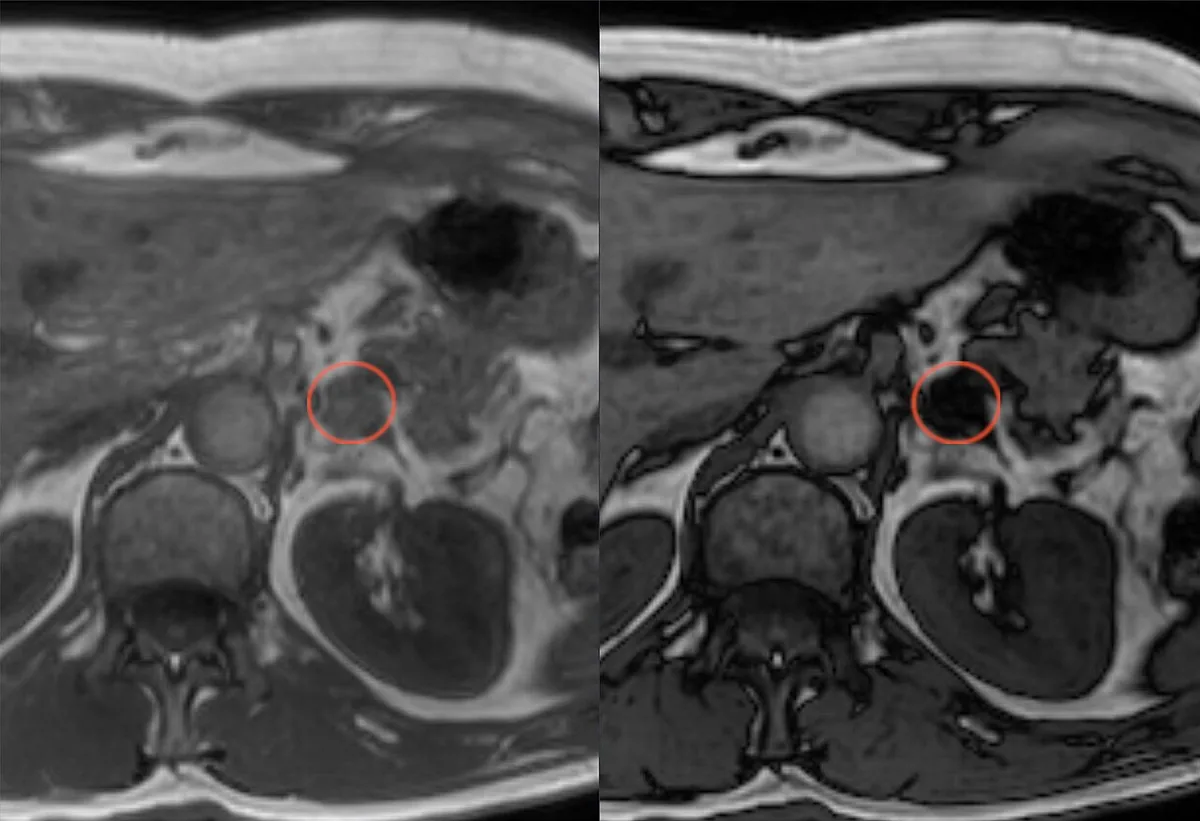 MRI
MRI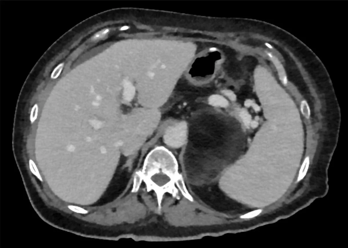 CT
CTDefinition
An adrenal incidentaloma is an adrenal mass discovered incidentally on imaging performed for another reason, such as abdominal CT or MRI. Every incidentaloma raises two questions: is the mass hormonally active, and is it benign or malignant? Most incidentalomas are non-functioning adrenocortical adenomas.
Classification
Causes
- Adrenal cortex: adenoma (non-functioning or functioning), nodular hyperplasia, adrenocortical carcinoma.
- Adrenal medulla: phaeochromocytoma, ganglioneuroma.
- Other tumors: myelolipoma, lipoma and others.
- Cysts and pseudocysts, hematomas and hemorrhage.
- Metastases, most often from lung cancer, also from breast, kidney and gastrointestinal cancers.
- Pseudo-adrenal masses arising from neighbouring organs.
- Bilateral masses: consider metastases, lymphoma, infections (e.g. tuberculosis, histoplasmosis) and hemorrhage.
Occurrence & epidemiology
Incidentalomas are found in about 1–5 % of cross-sectional imaging studies that include the adrenals and in about 2.3 % of autopsy series. Non-functioning adenomas account for about 70 %. In a large Japanese survey about 25 % were hormonally active: cortisol-producing 10.5 %, aldosterone-producing 5.1 %, catecholamine-producing 8.5 %. Adrenocortical carcinoma was found in 1.4 % and metastases in 3.7 %. With a known current or previous cancer outside the adrenal gland, the proportion of metastases rises to 50–75 %.
Clinical features
Most patients are asymptomatic. However, a significant proportion of apparently non-functioning adenomas secrete cortisol autonomously (mild autonomous cortisol secretion) without the classic Cushing picture; clues are weight gain, hypertension and diabetes mellitus. Cardiometabolic comorbidities are common with both non-functioning adenomas and mild cortisol autonomy.
Adrenal insufficiency is rare and occurs only with bilateral involvement. Massive bilateral hemorrhage presents with abdominal pain, a falling hematocrit and signs of acute adrenal insufficiency. A non-functioning adrenocortical carcinoma usually presents as an infiltrating retroperitoneal process or with metastases.
Diagnosis
Hormonal evaluation
- Cortisol autonomy: overnight dexamethasone suppression test; a serum cortisol of 50 nmol/l (1.8 µg/dl) or more the next morning is considered abnormal.
- Phaeochromocytoma: plasma free metanephrines or 24-hour urinary fractionated metanephrines. The dexamethasone suppression test is done only after phaeochromocytoma has been excluded, because it can provoke a hypertensive crisis in catecholamine-secreting tumors.
- Primary aldosteronism: plasma aldosterone and renin (aldosterone-to-renin ratio).
- Suspected adrenocortical carcinoma: additionally androgens and precursors such as DHEA-S, androstenedione, 17-OH progesterone and 11-deoxycortisol.
Imaging criteria
- Unenhanced CT: an attenuation of 10 HU or less indicates a lipid-rich, benign adenoma; this cut-off is used in current European guidelines.
- Guideline criteria for benignity: diameter below 4 cm and unenhanced attenuation of 10 HU or less.
- Features suggesting malignancy: diameter of 4 cm or more and unenhanced attenuation above 20 HU.
- CT with delayed contrast washout: an absolute washout above 60 % and a relative washout above 40 % suggest an adenoma.
- Chemical-shift MRI: signal loss on opposed-phase images indicates a lipid-rich adenoma.
Additional imaging notes
- Lipid-poor adenomas: at least 30 % of adenomas are lipid-poor and measure above 10 HU; their density then resembles phaeochromocytomas and malignant lesions.
- Size: in a prospective study 98 % of adrenocortical carcinomas measured at least 4 cm; none was smaller than 2 cm.
- Chemical-shift MRI: an alternative, particularly when iodinated contrast cannot be used.
- Fine-needle biopsy: only if metastasis or infection is suspected and only after phaeochromocytoma has been excluded; unsuitable when adrenocortical carcinoma is suspected because of the risk of tumor seeding.
Keep learning in the app
Further reading (open access)
Cross-references
Note: Learning content for medical education – not a treatment recommendation and no substitute for diagnosis or treatment decisions in individual cases. Treatment and management are deliberately not covered on this page.