ECG interpretation: the scheme step by step
Also in German (Deutsch)
You do not read an ECG “at a glance” – you read it in the same order every time. If you walk through the same ten steps on every tracing, you miss less, and at the end you can put the result into one clean sentence for the discharge letter and the handover. This guide shows the scheme with normal values from freely accessible sources, typical findings and five real ECGs to interpret yourself. Because many readers are preparing for exams in Germany, every step also gives the German wording.
Before step one: name, paper speed, calibration
First check that the ECG belongs to the right person and when it was recorded. Then two technical details without which every number can be wrong:
- Paper speed: at 25 mm/s one small square (1 mm) is 40 ms and one large square (5 mm) is 200 ms ECGpedia. Some machines record at 50 mm/s – then a small square is only 20 ms and all waves look twice as wide.
- Calibration: the standard is 10 mm/mV, so the calibration pulse at the start of each row is 10 mm high. If it is smaller (e.g. 5 mm/mV), all deflections appear half as tall ECGpedia.
The scheme at a glance
The table summarises adult normal values; the sections below explain each step. Where sources differ, both values are shown.
| Step | What you look at | Normal (adults) | Source |
|---|---|---|---|
| 1. Rhythm | P before every QRS? Regular intervals? | P before every QRS, P upright in I/II, negative in aVR | MSD |
| 2. Rate | 300 ÷ large squares, or QRS in 10 s × 6 | 60–100/min | ECGpedia |
| 3. Axis | main deflection in I, II, III (and aVL) | −30° to +90° (vertical to left axis type) | MSD · DocCheck |
| 4. Intervals | measure P, PR, QRS, QT, calculate QTc | P < 120 ms · PR 120–200 ms (MSD: 100–200) · QRS ≤ 100 ms · QTc ♂ < 450, ♀ < 460 ms (MSD: up to 450 / 460) | ECGpedia · MSD |
| 5. P wave | height and shape in II and V1 | < 120 ms, < 0.25 mV (MSD: P pulmonale above 2 mm) | ECGpedia · MSD |
| 6. QRS complex | width, Q waves, R-wave progression, voltage | narrow; Q waves in V1–V3 not normal | MSD |
| 7. ST segment | elevation or depression at the J point, which region? | isoelectric; elevation ≥ 1 mm in 2 contiguous leads (V2–V3 own cut-offs) = infarction criterion | MSD · Avdikos 2022 |
| 8. T and U wave | direction relative to QRS, inverted or peaked T, U wave | T concordant with QRS; small U wave possible | MSD |
| 9. Previous ECG | findings new or known? | new or known? | ECGpedia |
| 10. Summary | one-sentence conclusion | one-sentence conclusion | ECGpedia |
Practice ECGs: interpret them yourself: The five ECGs on this page are real recordings with a free licence. Walk through the scheme on each one before you open the answer. Rates are counted from the 10-second strips and are therefore approximate.
1Rhythm: sinus rhythm or not?
What you look at
Is there a P wave before every QRS complex and a QRS complex after every P wave? Are the intervals regular? Best judged on the long rhythm strip (usually lead II).
Normal values
- Sinus rhythm: a P wave before every QRS, constant PR interval; the P wave is upright in most leads and negative in aVR, P axis between 0° and 75° MSD
Common findings
- respiratory sinus arrhythmia (rate varies with breathing)
- atrial fibrillation (irregularly irregular, no P waves)
- atrial flutter (saw-tooth waves)
- supraventricular and ventricular ectopic beats
- AV nodal re-entrant tachycardia
- ventricular tachycardia
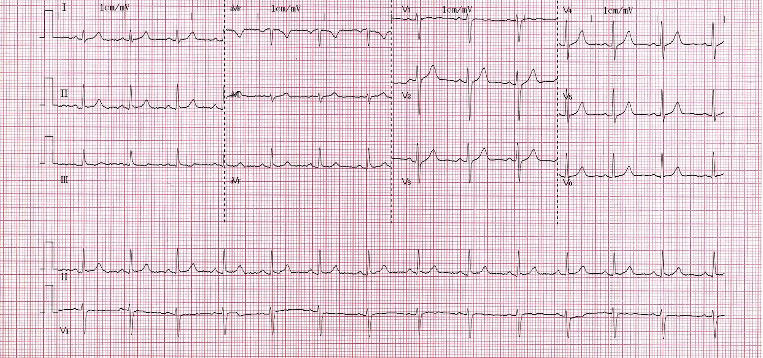
Interpret it yourself: rhythm, rate, axis, intervals, ST segment, T wave. Show answer
Sinus rhythm – a P wave before every QRS complex, P upright in I and II and negative in aVR, constant intervals. Rate about 80/min (14 QRS complexes in the 10-second rhythm strip × 6). Axis: QRS positive in I, II and III and taller in III than in I – in German terms a Steiltyp (+60° to +90°). Intervals normal, narrow QRS. No ST elevation or depression, T waves concordant with the QRS.
For comparison, the automatic measurement in the cropped header of the original: heart rate 82/min, PR 138 ms, QRS 91 ms, QT/QTc 344/404 ms, axis 70°. The machine classifies the ECG as “within normal limits”.
In German: „Sinusrhythmus, Herzfrequenz 82/min, Steiltyp, PQ 138 ms, QRS 91 ms, QTc 404 ms. Keine Erregungsausbreitungs- oder Erregungsrückbildungsstörungen.“
2Rate: normal, slow or fast
What you look at
With a regular rhythm: 300 divided by the number of large squares between two R waves (at 25 mm/s). With an irregular rhythm: count all QRS complexes in the 10-second strip and multiply by 6. At 50 mm/s a large square is only 100 ms – then use 600 divided by the number of large squares.
Normal values
- 60–100/min; below that bradycardia, above it tachycardia ECGpedia
Common findings
- sinus bradycardia, sinus tachycardia
- atrial fibrillation with slow, normal or rapid ventricular rate
- slow escape rhythm in AV block
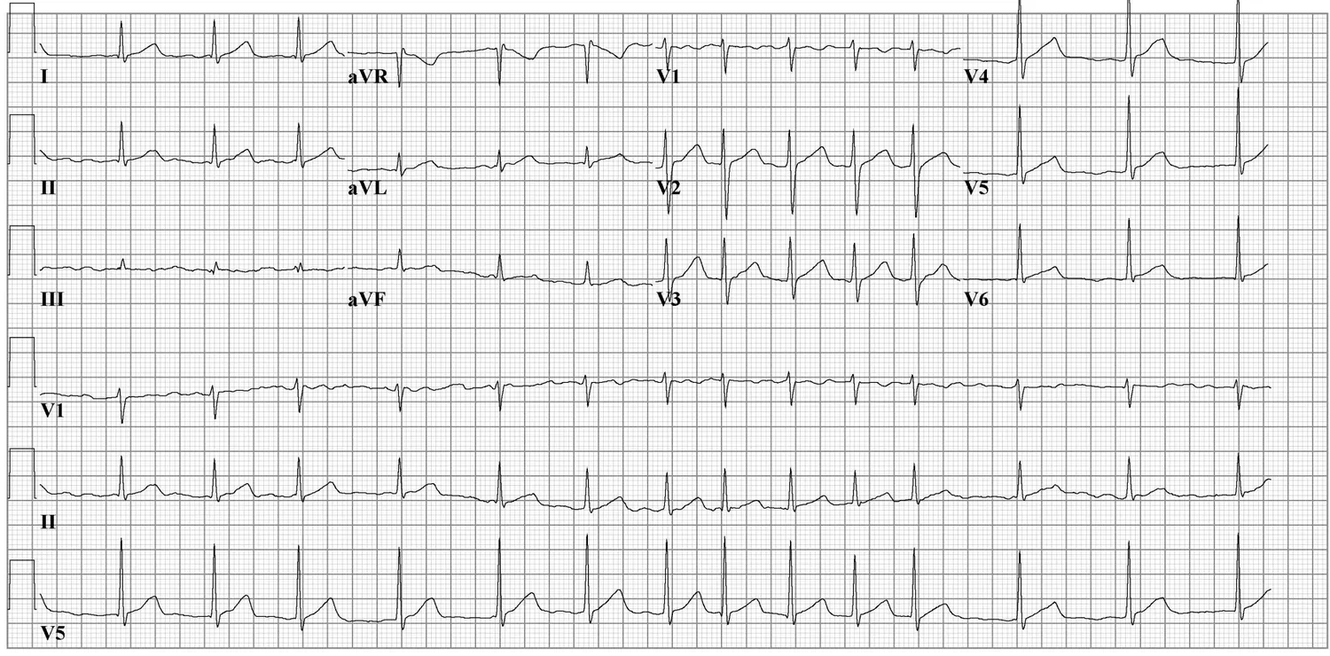
Interpret it yourself: is the rhythm regular? Can you see P waves? What is the ventricular rate? Show answer
Atrial fibrillation: the RR intervals vary constantly without any pattern (irregularly irregular) and there are no distinct P waves. Ventricular rate about 85/min (14 QRS complexes in the 10-second strip) – a normal ventricular rate. Narrow QRS complexes.
With an irregular rhythm the single-RR-interval method does not work; count the QRS complexes across the whole strip.
In German: „Vorhofflimmern mit normofrequenter Kammeraktion um 85/min, schmale QRS-Komplexe.“
3Axis: the electrical heart axis (German “Lagetyp”)
What you look at
Which way does the main QRS deflection point in leads I, II and III? A simple approach: QRS positive in I and II and negative in III means a left axis type (Linkstyp); all three positive means an intermediate or vertical axis – a taller R in I than in III points to the intermediate type (Indifferenztyp), a taller R in III to the vertical type (Steiltyp). If unsure, check aVL: positive in the intermediate type, negative in the vertical type.
Normal values
- Normal QRS axis: −30° to +90° MSD · ECGpedia
- German axis categories: überdrehter Linkstyp below −30°, Linkstyp −30° to +30°, Indifferenztyp +30° to +60°, Steiltyp +60° to +90°, Rechtstyp +90° to +120°, überdrehter Rechtstyp above +120° DocCheck
- Physiological in adults: vertical to left axis type; left axis type becomes more common with age DocCheck
Common findings MSD
- left axis deviation (−30° to −90°), e.g. in left anterior fascicular block (about −60°) or after inferior myocardial infarction
- right axis deviation (above +90°) – not the normal finding in adults
4Intervals: measuring P, PR, QRS and QT/QTc
What you look at
Measure in the lead where the waves are clearest. PR: from the start of the P wave to the start of the QRS complex. QRS: from start to end of the ventricular complex. QT: from the start of the QRS to the end of the T wave – it depends on heart rate and is therefore corrected (QTc), for example with Bazett’s formula.
Normal values
- P wave: under 120 ms ECGpedia
- PR interval: 120–200 ms according to ECGpedia; the MSD Manual gives 100–200 ms. Over 200 ms: first-degree AV block ECGpedia · MSD · MSD AV-Block
- QRS: 70–100 ms (MSD) or up to 100 ms (ECGpedia); 100–110 ms (MSD) or 110–119 ms (ECGpedia): incomplete bundle branch block or non-specific delay; 120 ms or more: complete bundle branch block or intraventricular conduction delay MSD · ECGpedia
- QTc: according to the MSD Manual 350–450 ms in men and 360–460 ms in women; according to ECGpedia prolonged from 450 ms (men) or 460 ms (women) and short at 390 ms or less. Asensio 2020 draws the limits differently: normal up to 430 ms in men and up to 450 ms in women, “borderline” up to 450 and 470 ms, prolonged from 460 and 480 ms; a QTc of 500 ms or more is described there as a risk marker MSD · ECGpedia · Asensio 2020
Common findings MSD AV-Block
- first-degree AV block (PR over 200 ms), second-degree AV block Mobitz I (Wenckebach: PR lengthens until a QRS is dropped) and Mobitz II (PR constant, a QRS is suddenly dropped), third-degree AV block (no conduction)
- short PR with delta wave (pre-excitation, WPW pattern)
- right and left bundle branch block (broad QRS)
- prolonged QTc (e.g. long QT syndrome, electrolyte disorders, drugs)
You can compare QTc formulas (Bazett, Fridericia, Framingham) with the QTc calculator.
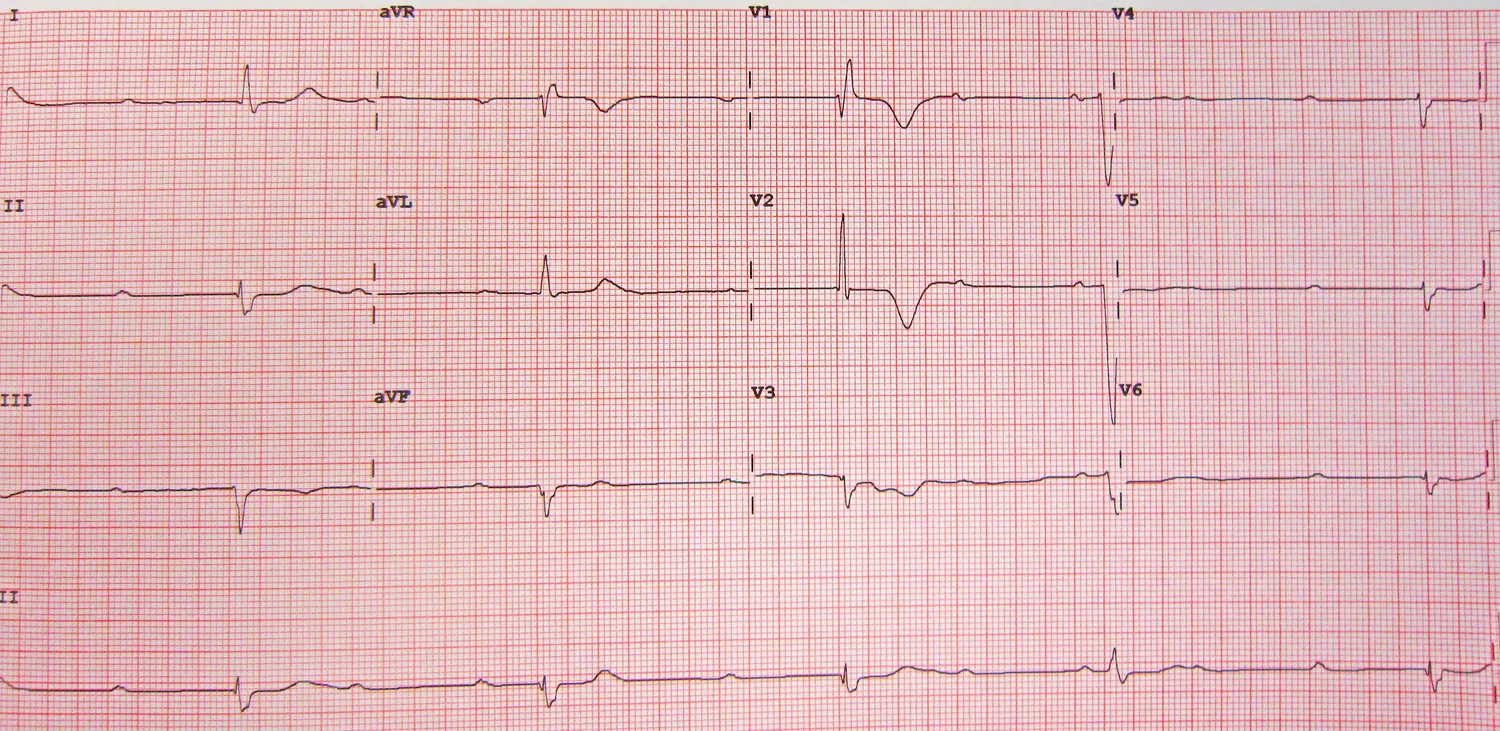
Interpret it yourself: how do the P waves relate to the QRS complexes? How long is the PR interval? Show answer
Third-degree (complete) AV block: the P waves are regular (atrial rate about 75/min), the QRS complexes are much less frequent (ventricular rate about 30/min) and have no fixed relation to the P waves – the distance between P and QRS changes from beat to beat. There is no meaningful PR interval: atria and ventricles beat independently (AV dissociation). In complete AV block no impulse is conducted; the ventricles are driven by an escape rhythm MSD AV-Block. One complex in rhythm strip II comes slightly early – whether it is conducted after all cannot be said with certainty from the image.
Emergency pattern: third-degree AV block with such a slow ventricular rate needs immediate medical assessment.
In German: „Totaler AV-Block (AV-Block III. Grades) mit bradykardem Ersatzrhythmus um 30/min, Vorhoffrequenz um 75/min.“
5P wave: shape and height
What you look at
Is the P wave in II small and rounded? Is it too tall or too wide, double-peaked, or markedly biphasic in V1?
Normal values
- duration under 120 ms, height under 0.25 mV (2.5 mm) ECGpedia
- The MSD Manual describes right atrial enlargement (P pulmonale) as a P wave over 2 mm in II, III and aVF – a slightly lower limit MSD
Common findings
- P pulmonale (tall, peaked P wave) with right atrial strain
- P mitrale (broad, notched P wave) with left atrial strain
- absent P waves in atrial fibrillation
6QRS complex: width, height, Q waves, R-wave progression
What you look at
Is the QRS narrow or broad? Are there prominent Q waves? Does the R wave grow from V1 to V5 (R-wave progression), and where does the complex change from mainly negative to mainly positive (transition)? Are the deflections unusually tall (a hint of hypertrophy, e.g. by the Sokolow-Lyon index) or unusually small (low voltage)?
Normal values
- QRS duration see step 4; 120 ms or more is complete bundle branch block MSD
- According to the MSD Manual, normal Q waves last less than 0.05 s – except in V1 to V3, where any Q wave is considered abnormal MSD
Common findings
- right bundle branch block (rSR’ in V1, broad S in I and V6) and left bundle branch block (broad, slurred R in I and V6)
- pathological Q waves as a sign of an old infarction
- signs of hypertrophy, poor R-wave progression, low voltage
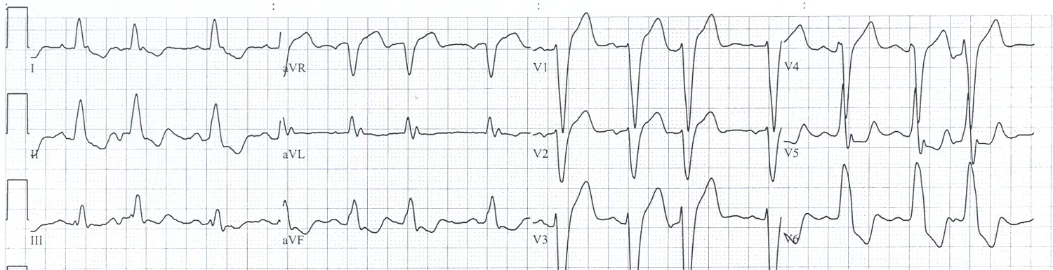
Interpret it yourself: is the QRS narrow or broad? What do V1 and V6 look like? How do the ST segment and T wave behave? Show answer
Left bundle branch block: the QRS complexes are broad (from 120 ms it counts as complete bundle branch block MSD). Deep, broad negative complexes in V1 to V3, broad slurred R waves in I and V6. ST segment and T wave point opposite to the main QRS deflection (discordant) – an expected consequence of the abnormal depolarisation in bundle branch block. According to the image description there are also occasional supraventricular ectopic beats.
Whether the block is new can only be decided by comparing with the previous ECG.
In German: „Kompletter Linksschenkelblock mit sekundären Erregungsrückbildungsstörungen, vereinzelte supraventrikuläre Extrasystolen.“
7ST segment: elevation or depression?
What you look at
Normally the ST segment runs at the level of the baseline (judged against the PR or TP segment). Check every lead: is the ST segment at the J point (end of the QRS) elevated or depressed – and does it affect contiguous leads of one region? The regions: anterior/septal V1–V4, inferior II, III, aVF, lateral I, aVL, V5–V6.
Normal values
- ST segment at baseline level or only slightly off it MSD
- According to the Fourth Universal Definition of MI (2018), the infarction criterion is new ST elevation at the J point in two contiguous leads of at least 1 mm; V2–V3 have their own cut-offs: ≥ 2 mm in men aged 40 or older, ≥ 2.5 mm in men under 40, ≥ 1.5 mm in women of any age – in each case without left ventricular hypertrophy or left bundle branch block Avdikos 2022 · UDMI 2018 · Saha 2025
- lead groups of the regions as above Saha 2025
Common findings
- ST-elevation myocardial infarction (STEMI) – emergency, immediate medical assessment
- ST depression (upsloping, horizontal, downsloping), also reciprocal to an elevation
- widespread ST elevation across several regions in pericarditis
- secondary ST-T changes in bundle branch block
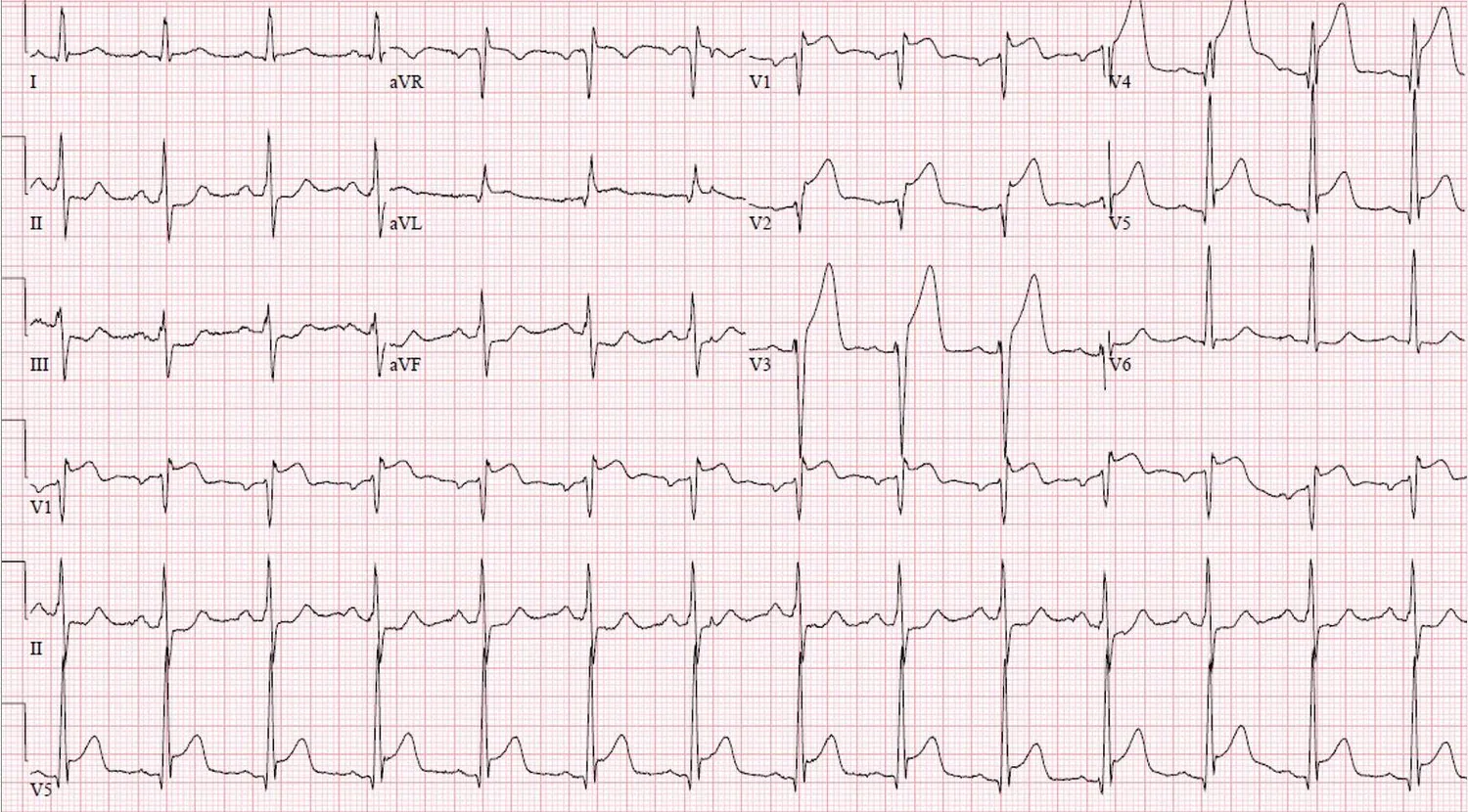
Interpret it yourself: in which leads is the ST segment elevated, where is it depressed? Which region fits? Show answer
Anterior ST-elevation myocardial infarction (STEMI): marked ST elevation in V1 to V5 (most pronounced in V2–V4, with tall T waves), less elevation in aVL; the authors of the case report also list I and V6. Reciprocal ST depression in II, III and aVF. Sinus rhythm at about 85/min. In the case report, cardiac catheterisation showed an occlusion of the left anterior descending artery (LAD, German RIVA) right at its origin.
Emergency: ST elevation in contiguous leads is an emergency pattern – immediate medical assessment.
In German: „Sinusrhythmus 85/min, ausgeprägte ST-Streckenhebungen in V1–V5 (betont V2–V4) und geringer in aVL mit spiegelbildlichen Senkungen in II, III und aVF – Bild eines akuten Vorderwandinfarkts (STEMI).“
8T wave and U wave
What you look at
Does the T wave point in the same direction as the main QRS deflection (concordant)? Are there negative T waves in contiguous leads? Is the T wave unusually tall and peaked? Is the T wave followed by a small U wave?
Normal values
- The T wave is normally concordant with the QRS complex; opposite (discordant) T waves can indicate an old or recent infarction MSD
- A U wave is often present in healthy people; it also occurs in hypokalaemia, hypomagnesaemia or ischaemia MSD
Common findings
- T-wave inversion (repolarisation abnormalities), e.g. in ischaemia, after infarction, in hypertrophy
- tall, peaked T waves in hyperkalaemia
- flattened T waves and prominent U waves in hypokalaemia
9Comparison with the previous ECG
What you look at
Is an older ECG available? Then compare every abnormal finding with it: new or known? A bundle branch block, T-wave inversion or Q wave that has been there for years means something different from the same finding appearing today. ECGpedia lists the comparison as its own step after the seven basic steps.
Normal values
- no normal value – what matters is the change ECGpedia
Common findings
- „im Vergleich zum Vor-EKG unverändert“ (unchanged compared with the previous ECG)
- „neu aufgetretener Linksschenkelblock“ (new left bundle branch block), „neu aufgetretene T-Negativierungen in V4–V6“ (new T-wave inversion in V4–V6)
10Summary: one sentence at the end
What you look at
The report ends with a one-sentence conclusion: what is normal, what is abnormal, and does it fit the question the ECG was recorded for? A normal ECG is called „Normalbefund“ or „altersentsprechend unauffällig“ (normal for age) in German. Emergency patterns such as ST elevation, third-degree AV block or ventricular tachycardia need immediate medical assessment – they belong in the first sentence.
Normal values
- ECGpedia ends the plan with the conclusion ECGpedia
How to word the report – in German
In a German discharge letter (Arztbrief) the ECG report is a short, noun-heavy text in a fixed order – the order of the scheme. In a handover you say the same in full sentences. The numbers in the examples are made up; the English line is a translation.
Model report for the discharge letter (normal ECG)
EKG: Sinusrhythmus, Herzfrequenz 72/min, Linkstyp. PQ 160 ms, QRS 90 ms, QT/QTc 380/416 ms. Regelrechte R-Progression, R/S-Umschlag in V3/V4. Keine ST-Streckenhebungen oder -senkungen, keine Erregungsrückbildungsstörungen. Im Vergleich zum Vor-EKG vom 03.02. unverändert.ECG: sinus rhythm, heart rate 72/min, left axis type (−30° to +30°). PR 160 ms, QRS 90 ms, QT/QTc 380/416 ms. Normal R-wave progression, transition in V3/V4. No ST elevation or depression, no repolarisation abnormalities. Unchanged compared with the previous ECG of 3 February.
Model report with an abnormal finding
EKG: Vorhofflimmern mit tachykarder Kammeraktion um 120/min, Indifferenztyp. QRS 90 ms, QTc nicht sicher bestimmbar. Keine ST-Streckenhebungen; deszendierende ST-Senkungen in V4–V6. Im Vergleich zum Vor-EKG vom 03.02. neu aufgetretenes Vorhofflimmern.ECG: atrial fibrillation with a rapid ventricular rate of about 120/min, intermediate axis (+30° to +60°). QRS 90 ms, QTc cannot be determined reliably. No ST elevation; downsloping ST depression in V4–V6. Atrial fibrillation is new compared with the previous ECG of 3 February.
In the handover (spoken)
„Im EKG zeigt sich ein Sinusrhythmus mit einer Frequenz von 72 pro Minute, Linkstyp. Die Zeiten sind normal, es gibt keine Hebungen und keine Senkungen. Im Vergleich zum Vor-EKG ist es unverändert.““The ECG shows sinus rhythm at 72 per minute, left axis type. The intervals are normal, there is no elevation and no depression. It is unchanged compared with the previous ECG.”
German building blocks you will need again and again
| German phrase | meaning |
|---|---|
| normofrequent / bradykard / tachykard | normal rate 60–100/min / below / above |
| regelmäßig / unregelmäßig, absolute Arrhythmie | regular / irregular, irregularly irregular |
| Linkstyp, Indifferenztyp, Steiltyp, Rechtstyp, überdrehter Links- oder Rechtstyp | German axis categories (see step 3) |
| PQ-Zeit verlängert („AV-Block I. Grades“) | prolonged PR interval (first-degree AV block) |
| schmale / breite QRS-Komplexe, kompletter / inkompletter Rechts- bzw. Linksschenkelblock | narrow / broad QRS, complete / incomplete right or left bundle branch block |
| regelrechte / verzögerte R-Progression, R/S-Umschlag in V3/V4 | normal / poor R-wave progression, transition in V3/V4 |
| ST-Streckenhebung / ST-Streckensenkung (aszendierend, horizontal, deszendierend) in … | ST elevation / ST depression (upsloping, horizontal, downsloping) in … |
| T-Negativierung in …, Erregungsrückbildungsstörungen (ERBS) | T-wave inversion in …, repolarisation abnormalities |
| im Vergleich zum Vor-EKG vom … unverändert / neu aufgetreten | unchanged / new compared with the previous ECG of … |
More on medical German: the discharge letter and doctor-to-doctor handover (both in German).
Common mistakes
- Starting with the most striking finding. If you stare at the ST elevation first, you miss the AV block. The scheme protects you.
- Trusting the machine blindly. The automatic interpretation is a help, not a report. In atrial fibrillation, artefacts or bundle branch block it often measures wrongly.
- Ignoring paper speed and calibration. At 50 mm/s or 5 mm/mV durations and heights no longer match the usual values.
- QT without rate correction. The QT interval depends on heart rate; what you assess is the QTc.
- Skipping the comparison. “New” or “known” decides what a bundle branch block, Q waves or T-wave inversion mean.
- Forgetting the summary. The report ends with one sentence naming the key findings – not just a list of numbers.
Practise ECGs with InnereFuchs
InnereFuchs (German) has an image trainer with ECGs, flashcards with spaced repetition and exam questions for internal medicine – free in the browser and as an app, with help in 13 languages for the FSP.
Open the web app App StoreFrequently asked questions
In which order do you interpret an ECG?
Always the same: rhythm, rate, axis, intervals (P, PR, QRS, QT/QTc), P wave, QRS complex, ST segment, T and U wave, comparison with the previous ECG and finally a summary. The fixed order makes sure nothing is missed – even when one finding jumps out at you.
How do you word an ECG report in a German discharge letter?
Briefly and in the order of the scheme, for example: „Sinusrhythmus, Herzfrequenz 72/min, Linkstyp, PQ 160 ms, QRS 90 ms, QTc 416 ms, keine ST-Streckenhebungen oder -senkungen, keine Erregungsrückbildungsstörungen, im Vergleich zum Vor-EKG unverändert.“ Abnormal findings are named with the leads, e.g. „T-Negativierung in V4–V6“ (T-wave inversion in V4–V6).
What are the normal values for PR, QRS and QTc?
For adults: PR 120–200 ms (ECGpedia; the MSD Manual gives 100–200 ms), QRS up to 100 ms, from 120 ms complete bundle branch block (MSD Manual), QTc up to 450 ms in men and up to 460 ms in women according to the MSD Manual and ECGpedia; other tables draw the limits slightly differently. The values are a guide, not rigid cut-offs.
Sources
Full references
- MSD: Cascino TM, Shea MJ: Electrocardiography. MSD Manual Professional Edition (full review 05/2026, updated 06/2026)
- MSD AV-Block: Mitchell LB: Atrioventrikulärer Block (atrioventricular block). MSD Manual, German professional edition (reviewed and updated 09/2024)
- ECGpedia: ECGpedia (CardioNetworks): Basics – normal ECG values, paper and calibration, the “7+2 step plan” for interpretation (accessed 13 Nov 2026)
- DocCheck: DocCheck Flexikon: Lagetyp (German axis categories with angle ranges and how to read them from leads I, II, III and aVL; German, as of 27 Aug 2026)
- Avdikos 2022: Avdikos G, Michas G, Smith SW: From Q/Non-Q Myocardial Infarction to STEMI/NSTEMI. Arch Acad Emerg Med 2022;10(1):e78 (open access; quotes the ST-elevation cut-offs of the Fourth Universal Definition of Myocardial Infarction 2018)
- UDMI 2018: Thygesen K, Alpert JS, Jaffe AS et al.: Fourth Universal Definition of Myocardial Infarction (2018). J Am Coll Cardiol 2018;72(18):2231–2264 (primary source of the ST cut-offs; cited here via Avdikos 2022)
- Saha 2025: Saha T, Soliman-Aboumarie H: Review of Current Management of Myocardial Infarction. J Clin Med 2025;14(17):6241 (open access; lead groups for infarct location)
- Asensio 2020: Asensio E, Acunzo R, Uribe W, Saad EB, Sáenz LC: Recommendations for the measurement of the QT interval during the use of drugs for COVID-19 infection treatment. J Interv Card Electrophysiol 2020;59(2):315 ff. (freely available via PMC; table of QTc normal values by sex)
Note: this guide explains how to describe an ECG in a structured way and names typical findings for learning. It is not a guide to diagnosis or treatment and does not replace a medical assessment. Normal values apply to adults and follow the sources listed (checked on 13 November 2026); machines, laboratories and textbooks may use other limits.