Hypokalemia
Board exam relevance: in 4 of 105 exam reports · rank 90- Synonyms
- low potassium, low blood potassium, potassium deficiency
- Specialty
- Internal medicine · Electrolytes & acid–base
- Images
- ECG 2
- Last updated
- 10/2026 · Dr. Pascal Bafteh
Contents
Images (2)
 ECG
ECG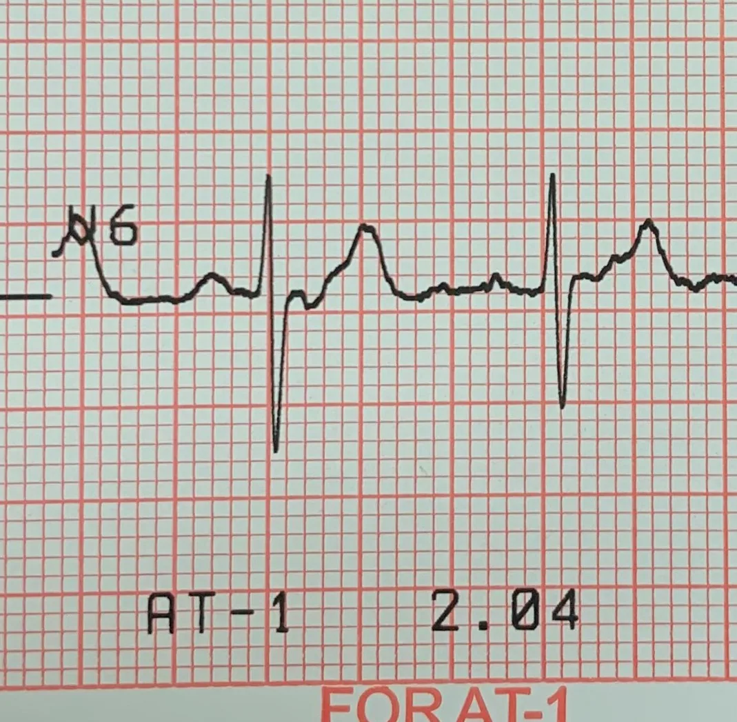 ECG
ECGDefinition
Hypokalemia is defined as a serum potassium concentration below 3.5 mmol/L. It results either from a deficit of total body potassium or from a shift of potassium into cells. The most common cause is increased loss via the kidneys or the gastrointestinal tract; low intake alone is rarely responsible.
Classification
- Mild (3.0 to 3.4 mmol/L): rarely causes symptoms.
- Moderate (2.5 to 3.0 mmol/L): usually causes muscle weakness and may lead to paralysis and respiratory failure.
- Severe (below 2.5 mmol/L): accompanied by ECG changes or severe symptoms.
Aetiopathogenesis
- Gastrointestinal losses: chronic diarrhea including laxative abuse, rarely a villous adenoma of the colon. In protracted vomiting or nasogastric suction, potassium is lost mainly via the kidneys, because metabolic alkalosis and volume-related aldosterone stimulation increase renal potassium excretion.
- Renal losses due to saluretics: by far the most common drug-related cause; surreptitious use of saluretics or laxatives is also a frequent cause of persistent hypokalemia, for example in people preoccupied with body weight.
- Aldosterone and mineralocorticoid excess: primary hyperaldosteronism, Cushing syndrome, renin-secreting tumors, congenital adrenal hyperplasia and liquorice intake (glycyrrhizin blocks the enzyme 11β-HSD, so that cortisol acts like aldosterone).
- Tubular disorders: Bartter and Gitelman syndromes (salt wasting with normal blood pressure), Liddle syndrome (with severe hypertension), renal tubular acidoses and Fanconi syndrome.
- Hypomagnesemia: a frequent accompaniment; it itself increases renal potassium losses.
- Shift into cells: increased insulin secretion (e.g. after glucose intake), beta-2-adrenergic stimulation, alkalosis, thyrotoxicosis (hypokalemic thyrotoxic periodic paralysis) and familial periodic paralysis, whose attacks are often precipitated by carbohydrate-rich meals or strenuous exercise.
Clinical features
- Muscles: weakness, cramps, fasciculations, tetany, respiratory muscle weakness up to respiratory failure and rhabdomyolysis.
- Gastrointestinal tract and circulation: paralytic ileus and hypotension.
- Kidney: persistent hypokalemia impairs urinary concentrating ability, causing polyuria with secondary polydipsia.
- Heart: premature beats, supraventricular and ventricular tachyarrhythmias, second- and third-degree AV block and eventually ventricular fibrillation. In pre-existing heart disease or with digitalis use, arrhythmias may occur even in mild hypokalemia.
Diagnosis
ECG
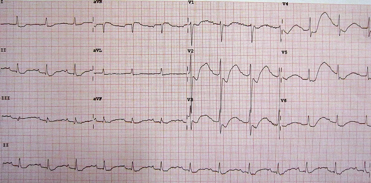
Cardiac effects are usually minor until potassium falls below about 3 mmol/L. Typical findings are ST-segment depression, flattening of the T wave and an increase in the U wave. As hypokalemia progresses, the T wave becomes smaller and the U wave larger; when the two merge, QT prolongation may be mimicked.
Laboratory tests and search for the cause
- Serum potassium and magnesium: the starting point of the work-up.
- History: the cause is usually evident from the history, particularly the drug history.
- 24-hour urinary potassium excretion: after excluding acidosis and other causes of intracellular shift. Values below 15 mmol/L suggest extrarenal (gastrointestinal) losses or low intake, values above 15 mmol/L renal losses.
- Blood pressure as a guide: renal potassium loss with hypertension points to an aldosterone-secreting tumor or Liddle syndrome; with normal blood pressure Bartter or Gitelman syndrome are possible, but hypomagnesemia, covert vomiting or surreptitious saluretic use are more common.
Keep learning in the app
Further reading (open access)
Cross-references
Note: Learning content for medical education – not a treatment recommendation and no substitute for diagnosis or treatment decisions in individual cases. Treatment and management are deliberately not covered on this page.