Urticarial vasculitis (normocomplementaemic and hypocomplementaemic)
- Synonyms
- Urtikaria-Vaskulitis, urtikarielle Vaskulitis, hypokomplementämische Urtikariavaskulitis, McDuffie-Syndrom, urticarial vasculitis, hypocomplementaemic urticarial vasculitis syndrome, HUVS, McDuffie syndrome
- Specialty
- Dermatology · Vascular skin disorders
- Images
- Clinical 1
- In the app
- 4 flashcards
- Last updated
- 10/2026 · Dr. Pascal Bafteh
Contents
Images (1)
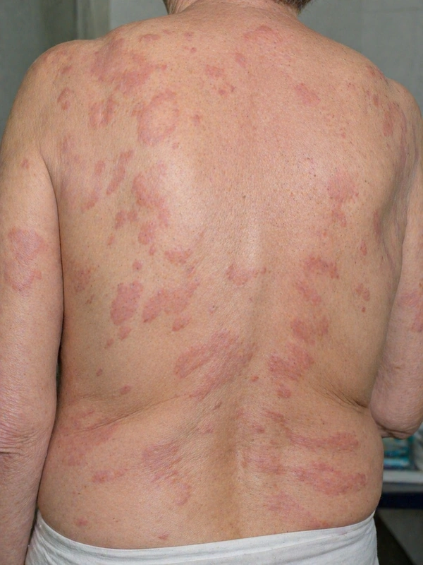 AI illustration
Definition
Urticarial vasculitis is a rare vasculitis of the small skin vessels. It produces urticaria-like weals that, unlike urticaria, usually last longer than 24 hours and heal with ecchymoses or hyperpigmentation; histology shows a vasculitis.
Classification
- Normocomplementaemic: normal complement levels, usually little or no systemic involvement.
- Hypocomplementaemic: low complement, associated with anti-C1q autoantibodies, more frequent extracutaneous symptoms and an association with systemic lupus erythematosus. The syndromic form with multi-organ involvement is called McDuffie syndrome.
Aetiopathogenesis
Urticarial vasculitis is regarded as immune complex-mediated: complement activation releases anaphylatoxins that attract neutrophils. Most cases are idiopathic. Associations include connective tissue diseases (SLE, Sjögren syndrome), IgM or IgA monoclonal gammopathies, leukaemia and internal cancers, viral infections (hepatitis B and C, infectious mononucleosis) and drug reactions.
Clinical features
- Weals or red patches and plaques, often burning or painful, sometimes itchy, occasionally with a pale centre and petechiae; individual lesions stay in place for more than 24 hours.
- Systemic features, especially in the hypocomplementaemic form: angioedema, joint pain (about 50%), fever, abdominal pain (about 20%), lymphadenopathy, photosensitivity, breathlessness, lung and kidney involvement.
Histology
In early lesions leukocytoclastic vasculitis with fibrinoid necrosis of dermal vessel walls and a neutrophil-rich perivascular infiltrate, in later lesions more lymphocytic; the typical findings are not present in every biopsy.
Diagnosis
- Skin biopsy to confirm the vasculitis.
- Complement levels to distinguish normo- from hypocomplementaemic disease; anti-C1q antibodies in hypocomplementaemia.
- Search for underlying disease and organ involvement, including renal function.
- Dermoscopy: purpuric patches and globules favour urticarial vasculitis over chronic urticaria.
Differential diagnoses
Keep learning in the app
References (selection)
Cross-references
Note: This page is intended for medical education and does not replace diagnosis or treatment decisions in individual cases.