Vitamin B12 deficiency and pernicious anemia
Board exam relevance: in 10 of 105 exam reports · rank 27- Synonyms
- cobalamin deficiency, B12 deficiency, pernicious anemia, Addison-Biermer anaemia, subacute combined degeneration, megaloblastic anaemia
- Specialty
- Internal medicine · Haematology & oncology
- Images
- Clinical 3 · Blood smear & cytology 1
- Last updated
- 10/2026 · Dr. Pascal Bafteh
Contents
Images (4)

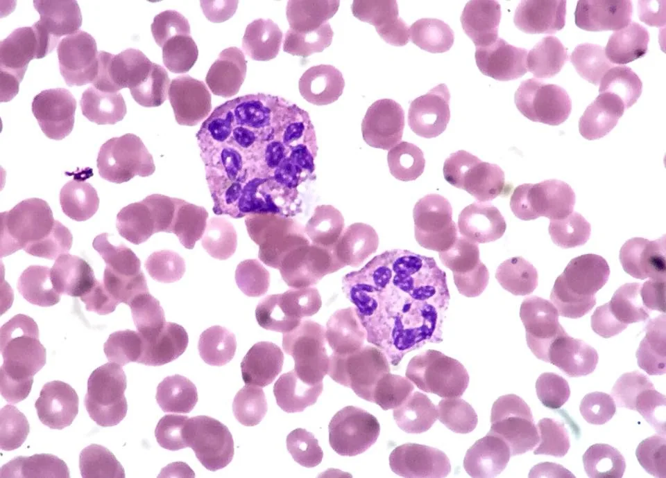 Blood smear & cytology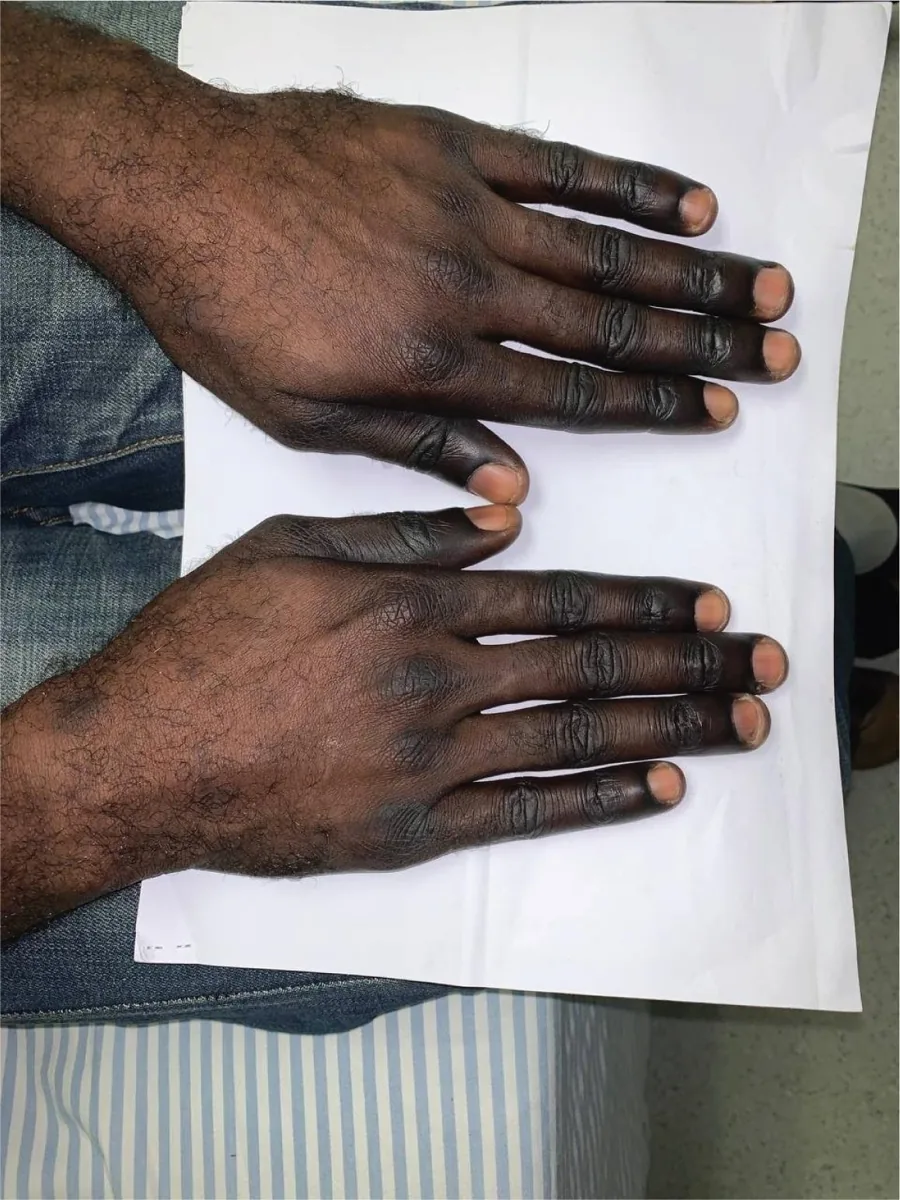

Definition
Vitamin B12 (cobalamin) deficiency causes megaloblastic, macrocytic anemia, damage to the white matter of the spinal cord and brain, and peripheral neuropathy. Cobalamin is needed for DNA synthesis, methylation reactions and the formation and maintenance of myelin.
Pernicious anemia is anemia due to B12 deficiency caused by autoimmune atrophic gastritis (type A gastritis) with loss of intrinsic factor. The term is often used synonymously with B12 deficiency, but strictly speaking it refers only to this cause.
Aetiopathogenesis
Physiology
B12 comes from animal foods (meat, offal, poultry, eggs, dairy products, fish and seafood). In the acidic gastric environment it is released from food and bound to haptocorrin (R protein). After cleavage by pancreatic enzymes in the small intestine it binds to intrinsic factor from the parietal cells of the gastric mucosa; the complex is absorbed in the terminal ileum. In plasma, transcobalamin II carries the vitamin to the tissues.
The liver stores large amounts of B12, which last about 3–5 years when intake stops; if enterohepatic recirculation is also interrupted, only months to about a year. The deficiency therefore usually develops very slowly.
Causes
- Impaired absorption (most common group): lack of intrinsic factor in autoimmune atrophic gastritis, destruction of the gastric mucosa, after surgery on the stomach (including bariatric procedures); reduced acid secretion in older age, so that food-bound B12 is not released; small-bowel disorders (Crohn disease involving the terminal ileum, celiac disease, tumors); chronic pancreatitis; consumption by bacterial overgrowth (blind loop syndrome) or fish tapeworm; advanced HIV infection; rarely inherited malabsorption (Imerslund-Gräsbeck syndrome).
- Inadequate intake: vegan diet, fad diets. Breastfed infants of vegan mothers can develop deficiency as early as 4–6 months of age because their liver stores are small.
- Impaired utilisation: enzyme defects, transport protein abnormalities, liver disease, repeated exposure to nitrous oxide.
Pernicious anemia and pathophysiology
In autoimmune atrophic gastritis, antibodies are directed against parietal cells and their components (intrinsic factor, H⁺/K⁺-ATPase). The results are hypochlorhydria, atrophy of the body and fundus and absent intrinsic factor. Hypochlorhydria leads via G-cell hyperplasia to high gastrin levels and to hyperplasia of enterochromaffin-like cells. The disease is associated with other autoimmune disorders: up to one third of people with autoimmune thyroiditis also have autoimmune gastritis. Younger women can also be affected.
B12 deficiency impairs DNA synthesis while RNA synthesis continues. Large cells with an immature nucleus (megaloblasts) arise; many precursors already die in the bone marrow (ineffective erythropoiesis). All cell lines are affected, so leukopenia and thrombocytopenia also develop later.
Clinical features
Hematological and gastrointestinal signs
- anemia develops insidiously and, because of adaptation, is often more severe than symptoms suggest: fatigue, weakness, pallor
- occasionally hepatomegaly and splenomegaly
- weight loss, poorly localised abdominal pain, diarrhea, loss of appetite
- glossitis with burning tongue
Neurological and psychiatric signs
Neurological disturbances develop independently of the anemia and can occur without anemia or macrocytosis; paraesthesias are sometimes the first sign. If they persist for a long time they can become permanent.
- Subacute combined degeneration of the dorsal columns and pyramidal tracts
- early: reduced position and vibration sense in the extremities, mild to moderate weakness, hyporeflexia, stocking-glove paraesthesias
- late: spasticity, extensor plantar responses, sensory ataxia with gait instability
- demyelinating or axonal peripheral neuropathy; touch, pain and temperature sensation are usually spared
- irritability, depressed mood, in advanced cases paranoia, delirium, confusion (sometimes mistaken for dementia) and orthostatic hypotension
Histology
Blood smear, bone marrow and gastric mucosa
- Smear: large oval red cells (macro-ovalocytes), anisocytosis and poikilocytosis, Howell-Jolly bodies (nuclear remnants); hypersegmented neutrophils appear early, neutropenia later; in severe cases thrombocytopenia with bizarrely shaped platelets.
- Bone marrow: megaloblastic erythropoiesis with maturation asynchrony – the cytoplasm is more mature than the nucleus; all cell lines show dyspoiesis.
- Gastric biopsy in pernicious anemia: atrophic mucosa of the body and fundus, sometimes with intestinal metaplasia.
Diagnosis
Laboratory tests
- Blood count: macrocytic anemia with MCV above 100 fl, high RDW, reticulocytopenia. Coexisting iron deficiency, thalassaemia trait or kidney disease can mask the macrocytosis; Howell-Jolly bodies and hypersegmentation nevertheless remain detectable.
- Signs of hemolysis due to ineffective erythropoiesis: LDH raised.
- Serum B12: values below 200 pg/ml (below 145 pmol/l) indicate deficiency; low-normal values just above this (up to about 300–350 pg/ml) are not diagnostic.
- Methylmalonic acid (MMA) and homocysteine: both raised in B12 deficiency, only homocysteine in folate deficiency. Renal failure also raises MMA.
- Holotranscobalamin (B12 bound to transcobalamin II): values below 40 pg/ml (below 30 pmol/l) indicate deficiency.
- Folate is always measured as well, because both deficiencies produce the same blood picture.
Establishing the cause
- Antibodies against intrinsic factor (specific for pernicious anemia) and against parietal cells (common but less specific); sensitivity and specificity are limited.
- Serum gastrin: often markedly raised in autoimmune gastritis (frequently above 1000 pg/ml).
- Gastroscopy with biopsy: confirms atrophic gastritis; autoimmune gastritis carries an approximately threefold increased risk of gastric adenocarcinoma, as well as of neuroendocrine tumors.
- Work-up for small-bowel and pancreatic disorders depending on the suspected cause; in autoimmune gastritis also a search for autoimmune thyroiditis.
Keep learning in the app
Further reading (open access)
Cross-references
More topics: Haematology & oncology
Note: Learning content for medical education – not a treatment recommendation and no substitute for diagnosis or treatment decisions in individual cases. Treatment and management are deliberately not covered on this page.