Non-Hodgkin lymphomas
Board exam relevance: in 12 of 105 exam reports · rank 20- Synonyms
- NHL, lymph node cancer, malignant lymphoma, B-cell lymphoma, DLBCL, follicular lymphoma, mantle cell lymphoma
- Specialty
- Internal medicine · Haematology & oncology
- Images
- CT 2 · Gross specimen 1 · Histology 2 · Blood smear & cytology 1 · Ultrasound 1
- Last updated
- 10/2026 · Dr. Pascal Bafteh
Contents
Images (7)
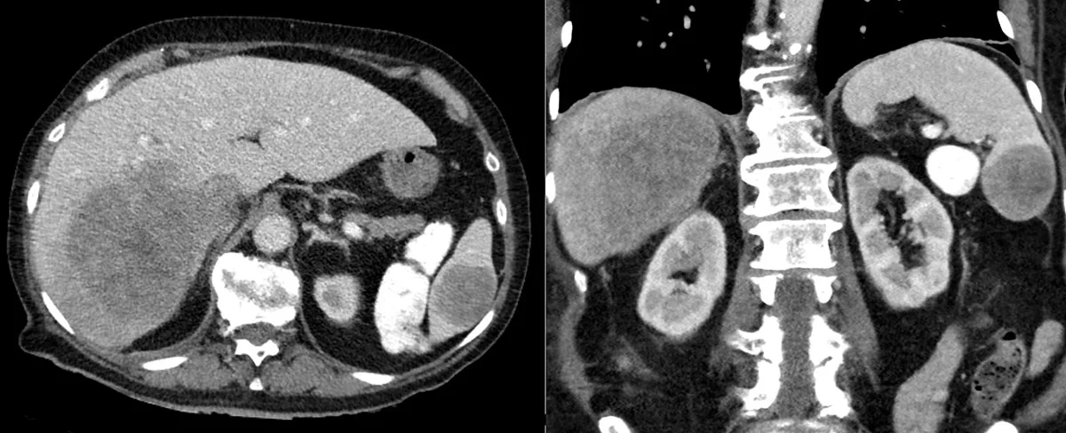 CT
CT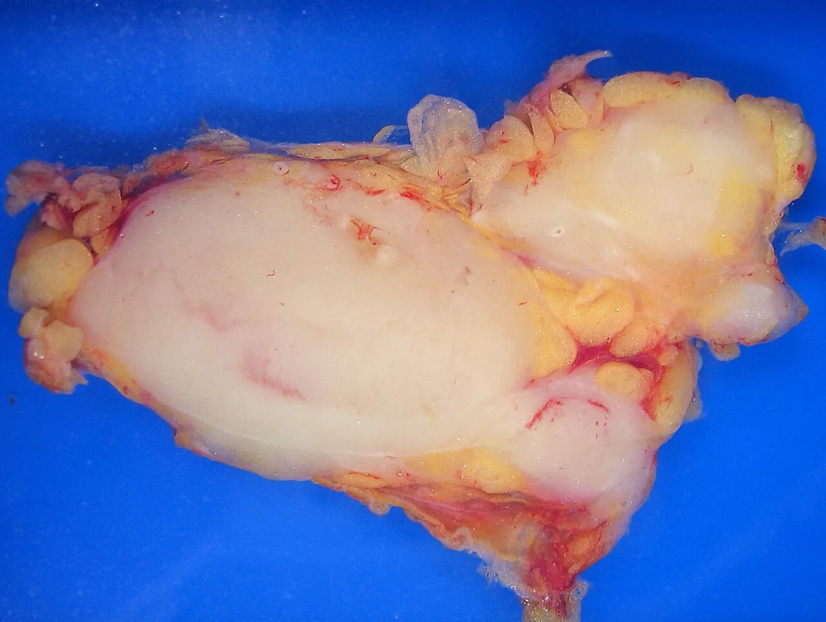 Gross specimen
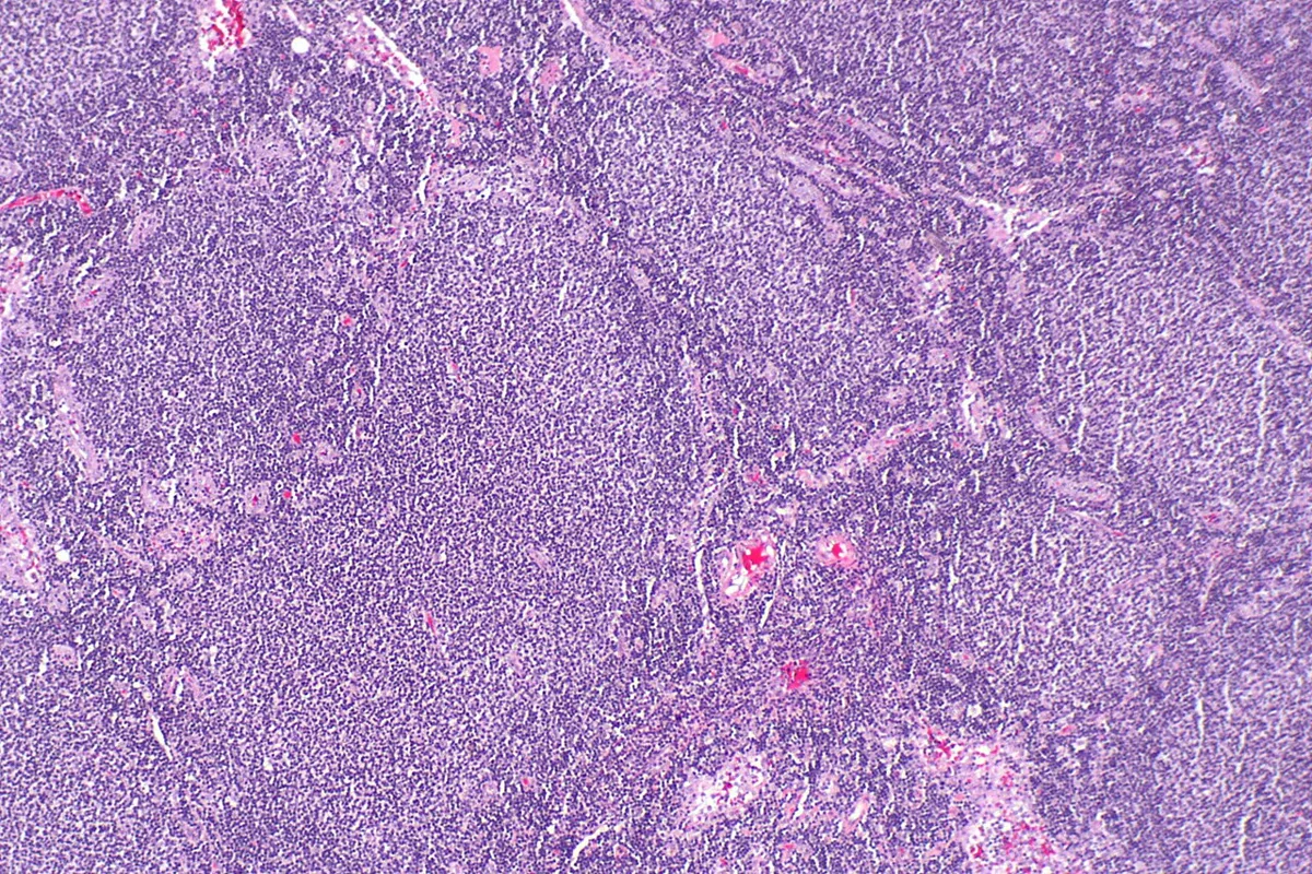 Histology
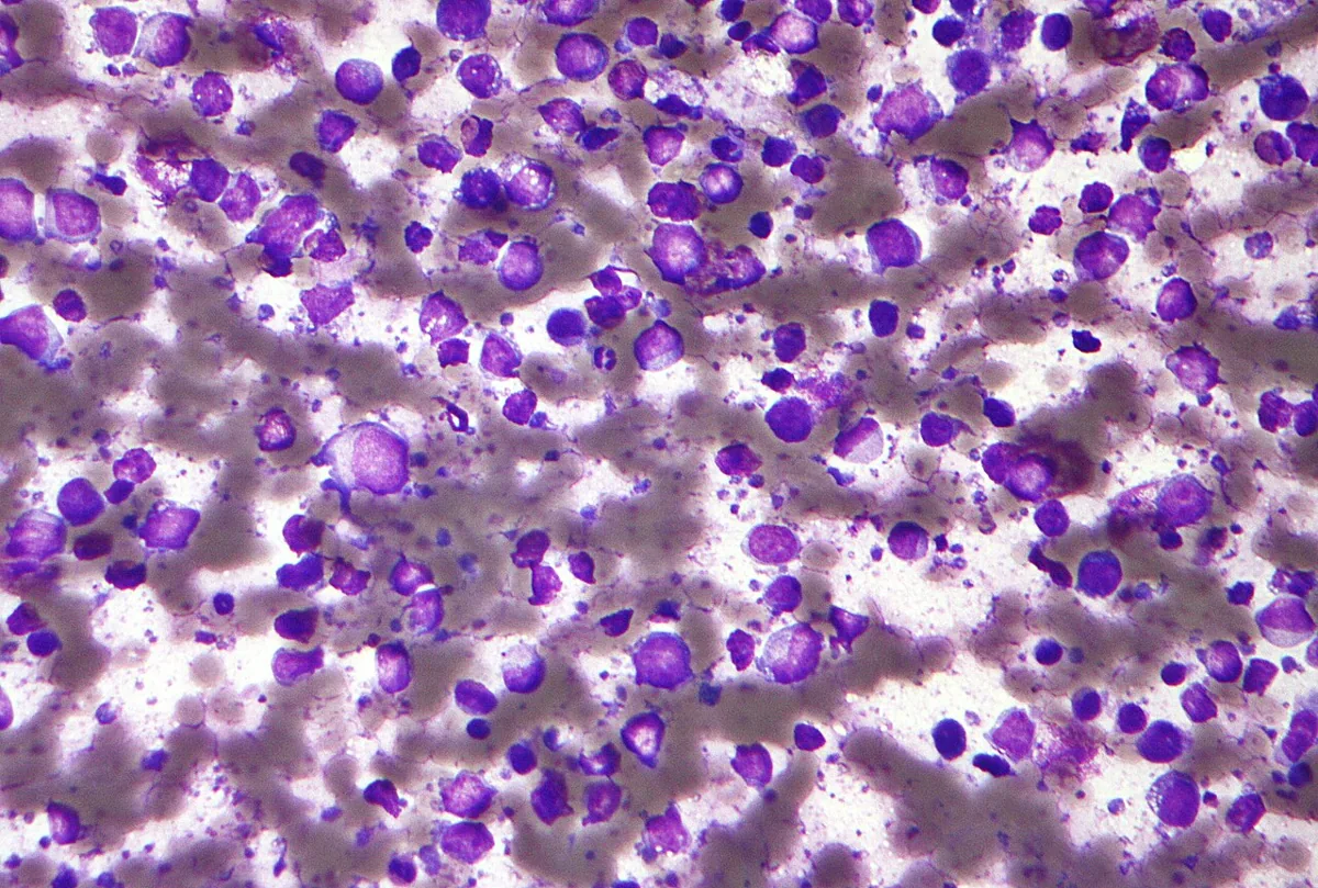 Blood smear & cytology
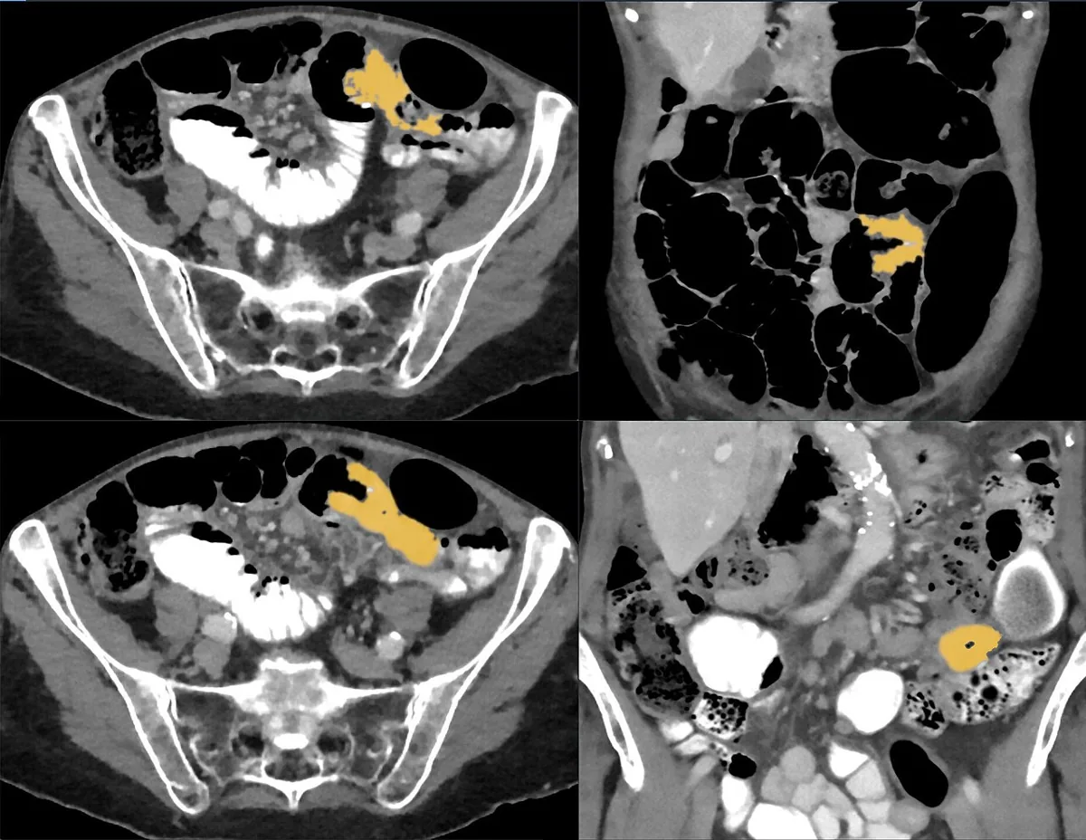 CT
CT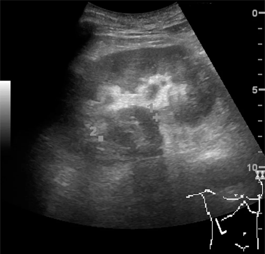 Ultrasound
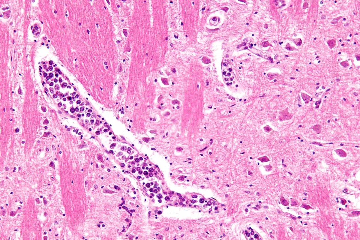 Histology
Definition
Non-Hodgkin lymphomas (NHL) are a heterogeneous group of malignant, monoclonal neoplasms of lymphoid cells. They arise in lymph nodes, bone marrow, spleen, liver, gastrointestinal tract, and other extranodal tissues. Most NHL arise from B lymphocytes, the rest from T lymphocytes or natural killer (NK) cells. The differentiation stage of the cell of origin shapes presentation and course. There is considerable overlap between lymphomas and leukemias, as lymphoma cells can also populate the blood and bone marrow.
Classification
Since 2022, two classifications have existed, the WHO classification (5th edition) and the International Consensus Classification (ICC). Both combine morphology, immunophenotype, genetics, and clinical picture. Clinically, the following division is common:
- Indolent lymphomas: slowly progressive
- Aggressive lymphomas: rapidly progressive
In children, NHL is almost always aggressive; follicular and other indolent lymphomas are unusual there.
Important entities
- Diffuse large B-cell lymphoma (DLBCL): most common entity among large B-cell lymphomas; aggressive
- Follicular lymphoma: predominantly (85 %) classic follicular lymphoma of centrocytes and centroblasts with translocation t(14;18) and IGH::BCL2 fusion; indolent
- Mantle cell lymphoma: IGH::CCND1 fusion from t(11;14) in ≥ 95 %, cyclin D1 overexpression
- Marginal zone lymphomas: extranodal (MALT lymphoma), nodal, and splenic; tumor cells usually CD5- and CD10-negative
- CLL/small lymphocytic lymphoma and lymphoplasmacytic lymphoma (Waldenström macroglobulinemia)
- Burkitt lymphoma: medium-sized cells with germinal center phenotype (CD10+, BCL6+), Ki-67 > 95 %, IG::MYC translocation; highly aggressive
- Hairy cell leukemia: BRAF V600E mutation in ≥ 95 %
- T-cell and NK-cell lymphomas, e.g., adult T-cell leukemia/lymphoma (HTLV-1-associated), anaplastic large cell lymphoma, cutaneous T-cell lymphomas
Staging
Staging follows the Lugano system (I–IV) as in Hodgkin lymphoma. At diagnosis, the disease is usually already disseminated.
Occurrence & epidemiology
NHL is considerably more common than Hodgkin lymphoma. In Germany, about 17,930 people were diagnosed with NHL in 2022 (about 8,100 women and 9,800 men). It is mainly a disease of older age: the mean age at diagnosis was 73 years in women and 71 years in men.
Aetiopathogenesis
The cause is usually unknown. Known risk factors:
- Viruses: human T-lymphotropic virus 1 (HTLV-1), Epstein-Barr virus (including endemic Burkitt lymphoma in Africa), hepatitis B and hepatitis C virus, HIV, human herpesvirus 8
- Helicobacter pylori: promotes gastric MALT lymphoma
- Congenital and acquired immunodeficiencies, e.g., immunosuppression in systemic rheumatic diseases or in solid organ transplant recipients; NHL is among the most common cancers in HIV infection
- Autoimmune disorders such as rheumatoid arthritis and Sjögren syndrome, chronic inflammation
- Chemicals: benzene, possibly certain herbicides and insecticides
- Genetic factors: first-degree relatives of patients with lymphoma have an increased risk
Clinical features
General symptoms
- Painless peripheral lymphadenopathy as the most common first symptom; nodes are rubbery, initially discrete, and later coalesce into masses. Usually several regions are involved.
- B symptoms (fever, night sweats, weight loss) and fatigue, especially in aggressive lymphomas; sometimes without palpable lymph nodes.
- Leukemic picture with lymphocytosis and bone marrow involvement in up to 50 % of children and about 20 % of adults with certain NHL.
- Anemia due to bleeding, hemolysis, hypersplenism, or bone marrow infiltration.
- Hypogammaglobulinemia in about 15 % at diagnosis, with increased susceptibility to infection.
Compression and extranodal involvement
Enlarged mediastinal and retroperitoneal lymph nodes cause compression symptoms:
- Superior vena cava: shortness of breath and facial edema (superior vena cava syndrome)
- Biliary tree: jaundice
- Ureters: hydronephrosis
- Bowel: vomiting and obstipation up to bowel obstruction
- Lymph drainage: chylous pleural or peritoneal effusion, lymphedema of a leg
Extranodal manifestations: skin (in B-cell lymphomas, erythematous nodules on the scalp or legs; in cutaneous T-cell lymphomas, erythema, papules, plaques, or tumors), gastrointestinal tract (particularly the terminal ileum in aggressive childhood lymphomas), meninges, testes. Adult T-cell leukemia/lymphoma runs a fulminant course with skin infiltrates, hepatosplenomegaly, and frequently hypercalcemia.
Histology
Histology shows destruction of the normal lymph node architecture with infiltration of the capsule and surrounding fat by neoplastic cells. Classification is made by immunophenotyping, demonstration of B- or T-cell clonality, cytogenetics, and, if applicable, gene sequencing.
Diagnosis
Tissue diagnosis
- Lymph node biopsy as the basis of diagnosis: open biopsy for an easily palpable node, CT- or ultrasound-guided core needle biopsy for involvement in the chest or abdomen; fine-needle aspiration often yields too little tissue.
- Review by hematopathologists experienced in lymphoma diagnosis; cytogenetics and flow cytometry require fresh tissue.
Staging and imaging
- FDG-PET/CT of the chest, abdomen, and pelvis; alternatively contrast-enhanced CT.
- Unilateral bone marrow aspiration and biopsy often, especially when the result changes classification.
- MRI of the brain and/or spinal cord in patients with neurologic symptoms.
Laboratory tests
- Complete blood count with differential (anemia, lymphocytosis), immunoglobulins.
- Serology for HIV, hepatitis B, and hepatitis C; in adult T-cell leukemia/lymphoma, additionally HTLV-1.
Keep learning in the app
Further reading (open access)
Cross-references
More topics: Haematology & oncology
Note: Learning content for medical education – not a treatment recommendation and no substitute for diagnosis or treatment decisions in individual cases. Treatment and management are deliberately not covered on this page.