Autoimmune hemolytic anemia (AIHA)
Board exam relevance: in 5 of 105 exam reports · rank 69- Synonyms
- warm autoimmune haemolytic anaemia, cold agglutinin disease, autoimmune hemolytic anemia, Coombs-positive anaemia
- Specialty
- Internal medicine · Haematology & oncology
- Images
- Blood smear & cytology 4
- Last updated
- 10/2026 · Dr. Pascal Bafteh
Contents
Images (4)
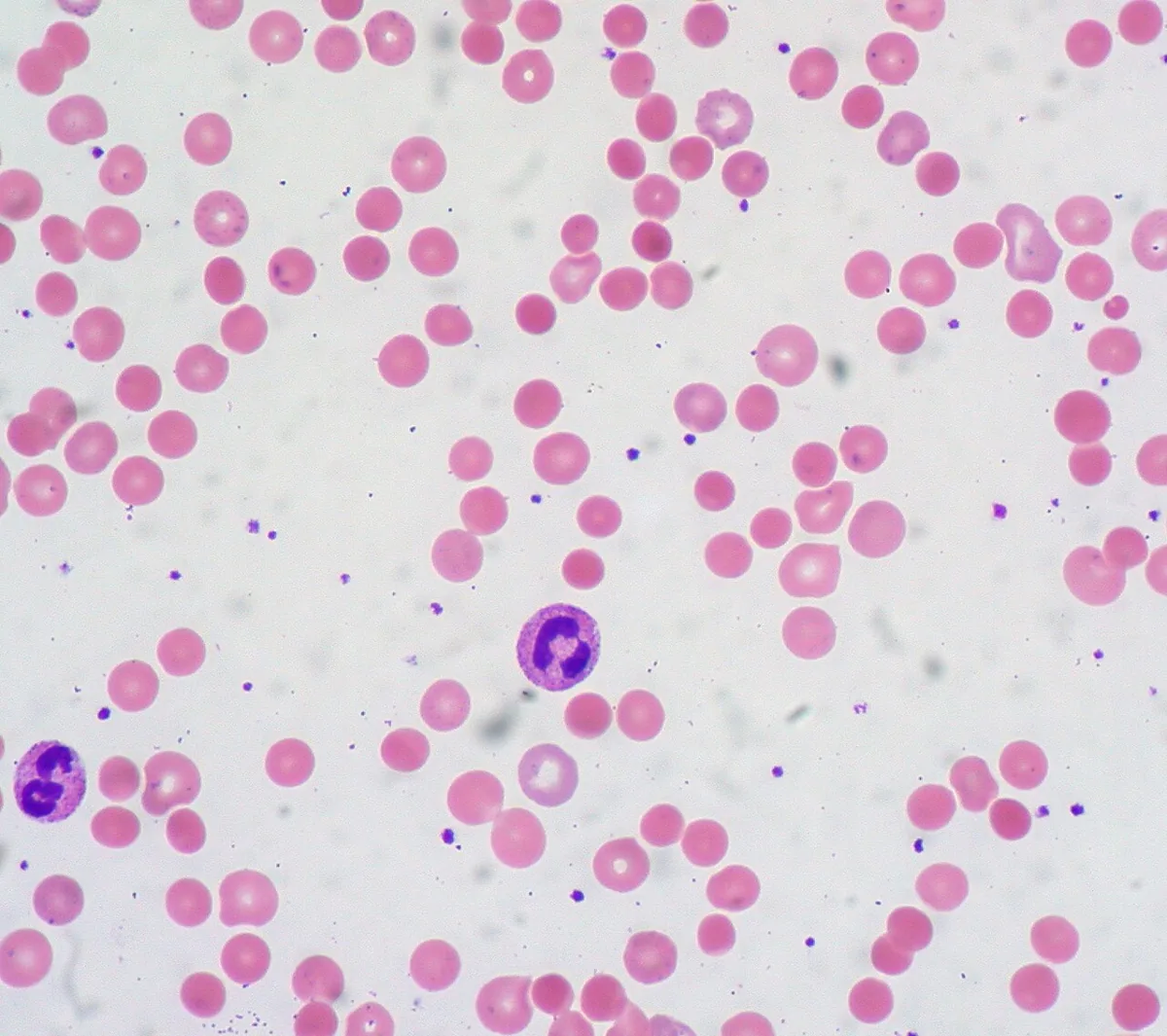 Blood smear & cytology
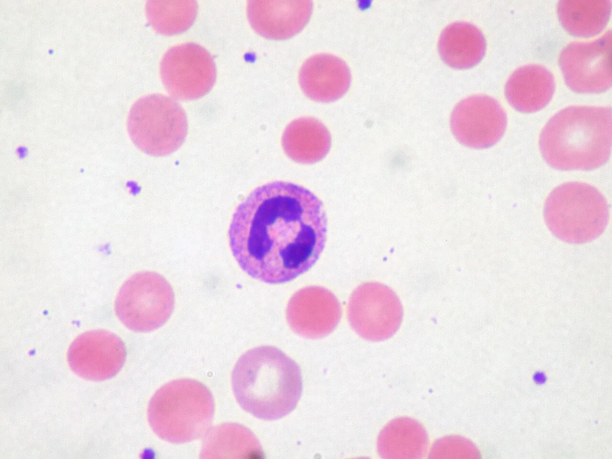 Blood smear & cytology
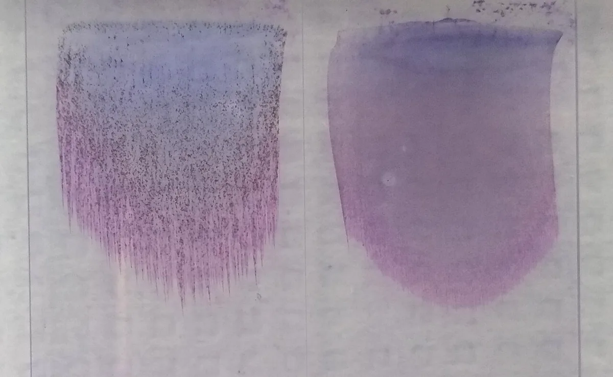 Blood smear & cytology
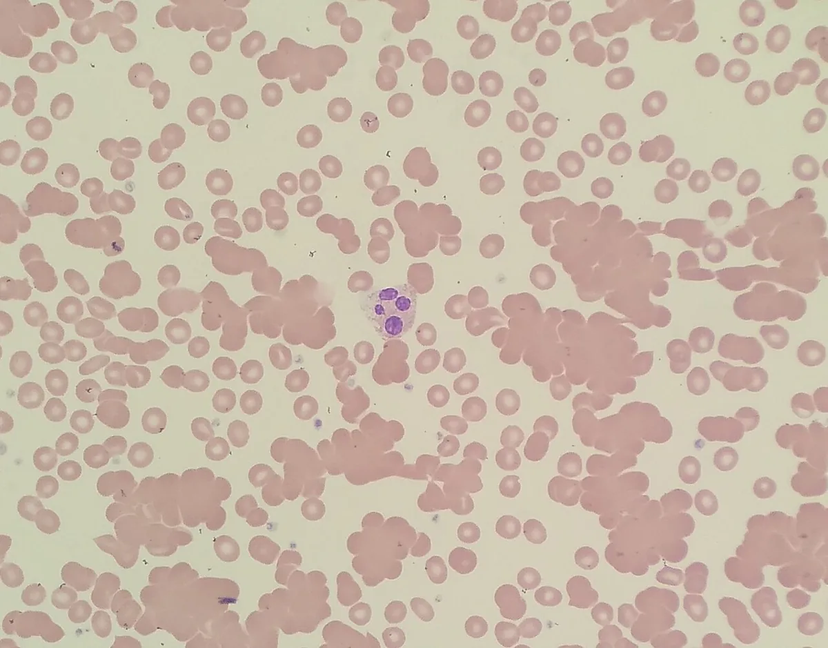 Blood smear & cytology
Definition
Autoimmune hemolytic anemia (AIHA) is an acquired hemolysis caused by autoantibodies against the body's own red cells. According to the temperature at which the antibodies bind best, warm and cold antibodies are distinguished. Hemolysis is predominantly extravascular; the direct antiglobulin test (Coombs test) confirms the diagnosis.
Classification
- Warm autoimmune hemolytic anemia (wAIHA): antibodies react at 37 °C, mostly polyclonal IgG against common red cell antigens (panagglutinins); destruction mainly in the spleen. Primary (idiopathic) or secondary, e.g. in systemic lupus erythematosus, lymphoma, chronic lymphocytic leukemia or drug-induced.
- Cold agglutinin disease (CAD): almost always clonal IgM against the I antigen, reacting below 37 °C; thermal amplitude matters more than titre. The primary form is usually associated with a clonal B-cell population (low-grade lymphoproliferative disorder), chronic and mainly seen in older adults.
- Secondary cold agglutinin syndrome: acute after infections, especially mycoplasma pneumonia (anti-I) or infectious mononucleosis (anti-i), or with overt lymphoma.
- Paroxysmal cold hemoglobinuria (Donath-Landsteiner): rare, mainly in children, usually after viral infections; an IgG against the P antigen binds in the cold and triggers intravascular hemolysis after rewarming.
- Mixed AIHA: warm and cold antibodies together.
Occurrence & epidemiology
The incidence of wAIHA is estimated at 1:50,000 to 1:100,000 and that of cold agglutinin disease at about 1:1,000,000 per year. wAIHA accounts for about 48–70 % and cold agglutinin disease for about 15–25 % of all AIHA; women are slightly more often affected by wAIHA. About half of wAIHA cases in adults are secondary. AIHA occurs in about 10 % of people with systemic lupus erythematosus and in 5–10 % of people with chronic lymphocytic leukemia.
Aetiopathogenesis
IgG-coated red cells are recognized and destroyed or partially phagocytosed by splenic macrophages; they lose membrane and become microspherocytes. IgM cold agglutinins bind in cooler body regions, activate complement and lead to C3 coating; the cells are mainly destroyed in the liver and spleen. In complement-amplifying situations such as infections, hemolysis can also be intravascular.
Drug-induced immune hemolysis arises through different mechanisms: formation of true autoantibodies against Rh antigens or antibodies against a complex of drug and red cell membrane (hapten mechanism).
Clinical features
- wAIHA: symptoms of anemia, jaundice, typically mild splenomegaly; in severe cases fever, chest pain, syncope, liver or heart failure. Venous thromboembolism is common.
- Cold agglutinin disease: acute or chronic hemolysis, usually not severe; cold-induced acrocyanosis and Raynaud phenomenon, increased thrombotic risk.
- Paroxysmal cold hemoglobinuria: after cold exposure severe back and leg pain, headache, vomiting, diarrhea, fever and dark brown urine; sometimes hepatosplenomegaly.
Histology
Blood smear
In wAIHA microspherocytes and polychromasia with a high reticulocyte count, few or no schistocytes. In cold agglutinin disease, red cells visibly clump on the smear when the blood is not warmed.
Diagnosis
- Hemolysis markers: reticulocytes, LDH and indirect bilirubin raised, haptoglobin low. With cold agglutinins, clumping in the analyser produces a spuriously high MCV and falsely low Hb; warming the sample largely normalises the values.
- Direct antiglobulin test: pattern IgG alone or IgG plus C3 in wAIHA; C3 without IgG in cold agglutinin disease and paroxysmal cold hemoglobinuria. About 5 % of AIHA cases are DAT-negative (very low antibody density, rarely IgA). A positive DAT without hemolysis occurs, e.g. with paraproteins, after immunoglobulin products or due to alloantibodies after donor blood.
- Indirect antiglobulin test: free antibodies in serum; positive with a negative DAT suggests alloantibodies rather.
- Cold agglutinin titre (at 4 °C; usually at least 1:64) and thermal amplitude (clinically relevant if binding occurs above 28–30 °C).
- Donath-Landsteiner test: specific for paroxysmal cold hemoglobinuria.
- Search for underlying disorders (secondary AIHA): ANA, antiphospholipid antibodies and complement if autoimmune disease is suspected; serum protein electrophoresis, immunofixation, B-lymphocyte immunophenotyping and imaging if lymphoma is suspected; immunoglobulin levels if immunodeficiency is suspected; serology for HIV, hepatitis B and C, CMV, EBV, parvovirus B19 and mycoplasma.
Keep learning in the app
Further reading (open access)
Cross-references
More topics: Haematology & oncology
Note: Learning content for medical education – not a treatment recommendation and no substitute for diagnosis or treatment decisions in individual cases. Treatment and management are deliberately not covered on this page.