Multiple myeloma
Board exam relevance: in 9 of 105 exam reports · rank 32- Synonyms
- plasma cell myeloma, myeloma, Kahler disease, bone marrow cancer, smoldering myeloma
- Specialty
- Internal medicine · Haematology & oncology
- Images
- Blood smear & cytology 2 · X-ray 4 · MRI 1 · Histology 1
- Last updated
- 10/2026 · Dr. Pascal Bafteh
Contents
Images (8)
 Blood smear & cytology
 X-ray
X-ray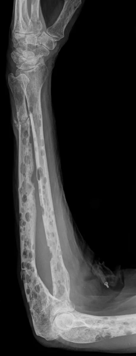 X-ray
X-ray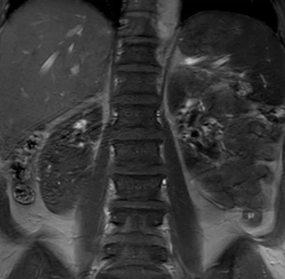 MRI
MRI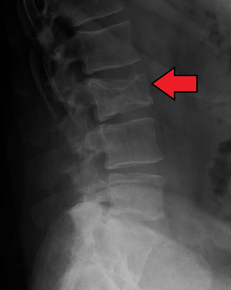 X-ray
X-ray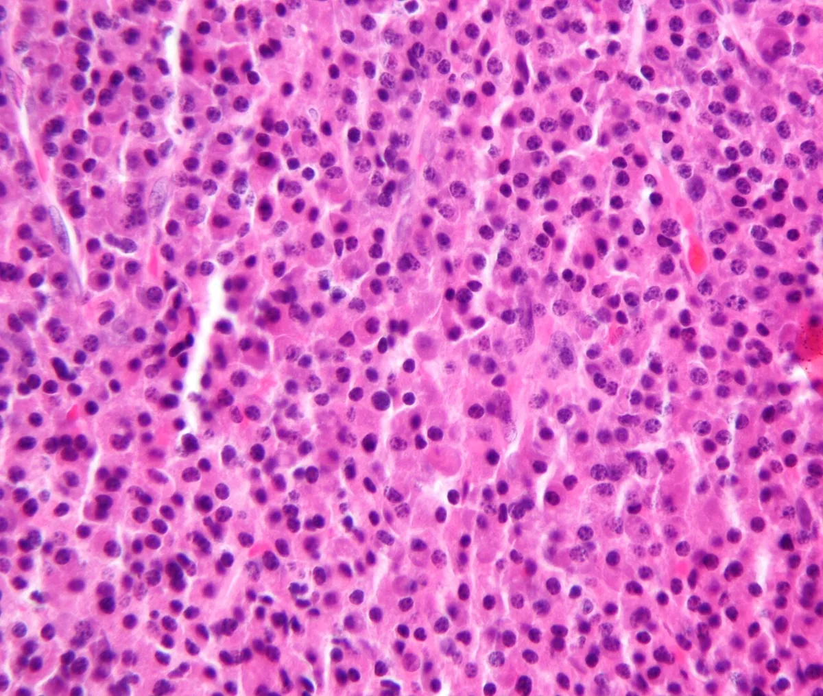 Histology
 X-ray
X-ray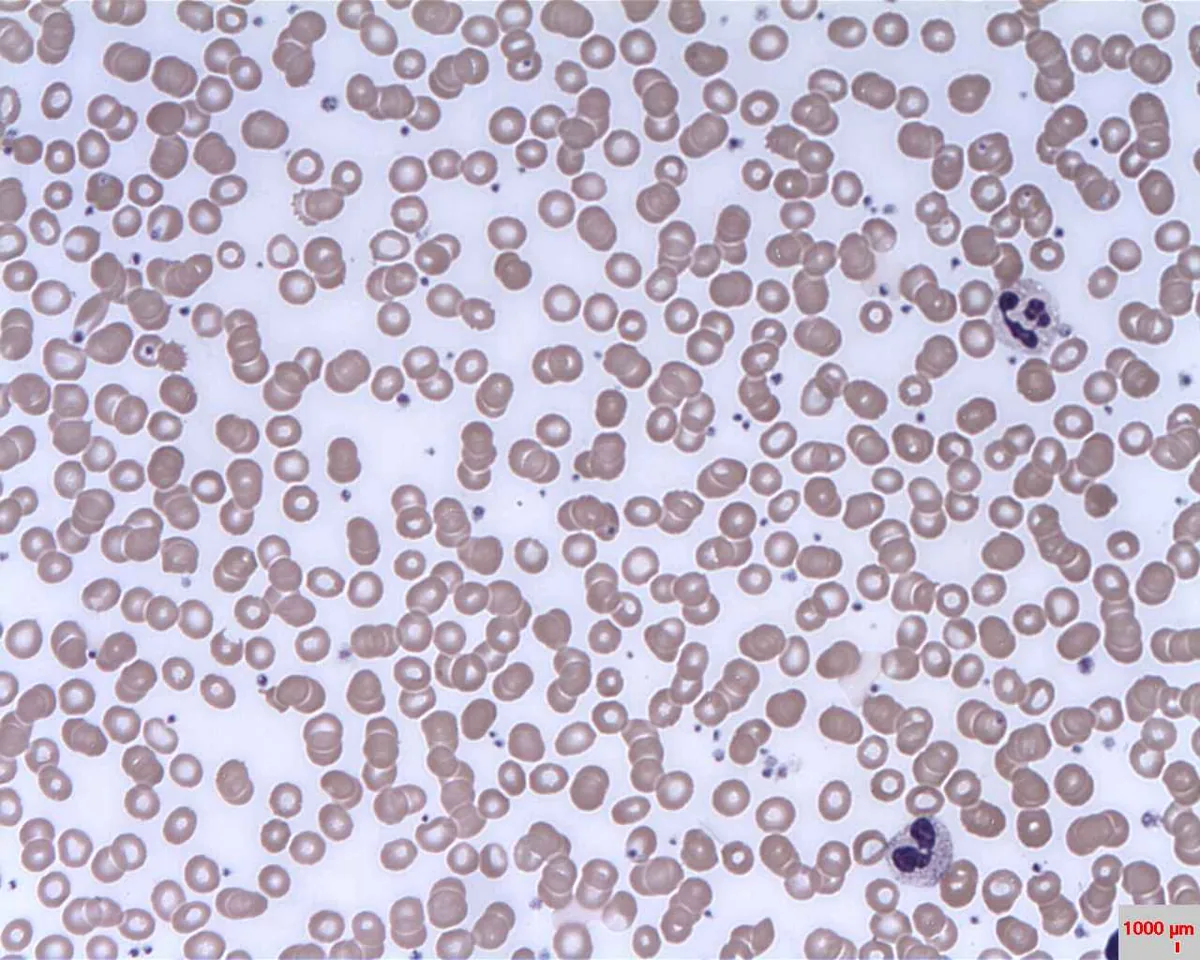 Blood smear & cytology
Definition
Multiple myeloma (synonym: plasmacytoma; in the WHO classification plasma cell myeloma) is a malignant proliferation of antibody-producing plasma cells. The clonal plasma cells produce a monoclonal immunoglobulin or its light chains (M-protein, paraprotein), usually grow multifocally in the bone marrow, and destroy adjacent bone. Typical consequences are bone pain and fractures, renal insufficiency, hypercalcemia, anemia, and susceptibility to infection. Virtually all myelomas arise from monoclonal gammopathy of undetermined significance (MGUS).
Classification
Diagnostic criteria
According to the International Myeloma Working Group (IMWG) criteria, myeloma is present with ≥ 10 % clonal plasma cells in the bone marrow or a plasmacytoma plus at least one myeloma-defining event:
- CRAB criteria: hypercalcemia (serum calcium > 0.25 mmol/L above normal or > 2.75 mmol/L), renal insufficiency (creatinine clearance < 40 mL/min or serum creatinine > 2 mg/dL), anemia (hemoglobin > 2 g/dL below normal or < 10 g/dL), osteolytic bone lesions on radiography, CT, or PET-CT
- SLiM criteria: ≥ 60 % clonal plasma cells in the bone marrow, involved/uninvolved free light chain ratio ≥ 100, more than one focal lesion (≥ 5 mm) on MRI
Smoldering myeloma: M-protein ≥ 3 g/dL and/or 10–59 % clonal plasma cells without a myeloma-defining event.
Types and staging
- By isotype: IgG in about 50–55 %, IgA in about 20 %, pure light chain myeloma (Bence Jones protein only) in 15–20 %, IgD and IgM 1–2 % each, IgE exceedingly rare; nonsecretory myeloma rare.
- Variants: extramedullary plasmacytoma, solitary plasmacytoma of bone, osteosclerotic myeloma (POEMS syndrome: polyneuropathy, organomegaly, endocrinopathy, M-protein, skin changes).
- Revised International Staging System (R-ISS): stage I with beta-2 microglobulin < 3.5 mg/L, albumin ≥ 3.5 g/dL, normal LDH, and standard-risk cytogenetics; stage III with beta-2 microglobulin ≥ 5.5 mg/L and high-risk cytogenetics [t(4;14), t(14;16), del(17p)] and/or elevated LDH; stage II all others.
Occurrence & epidemiology
In Germany, about 2,730 women and 3,790 men were newly diagnosed in 2022. Risk rises markedly with older age; the disease is extremely rare before age 45. The median age at diagnosis is about 70 years. People of African ancestry are affected about twice as often; men are affected slightly more often.
Aetiopathogenesis
- Risk factors: preexisting MGUS, older age, male sex, African ancestry, familial clustering; associations with HIV and hepatitis C infection; after intensive occupational benzene exposure, myeloma is recognized in Germany as an occupational disease under certain conditions.
- Genetics: hyperdiploidy (trisomies of odd-numbered chromosomes) in 50–60 %; translocations at the immunoglobulin heavy chain locus, e.g., t(11;14), t(4;14), t(14;16); high-risk features are t(4;14), t(14;16), del(17p), and 1q gain.
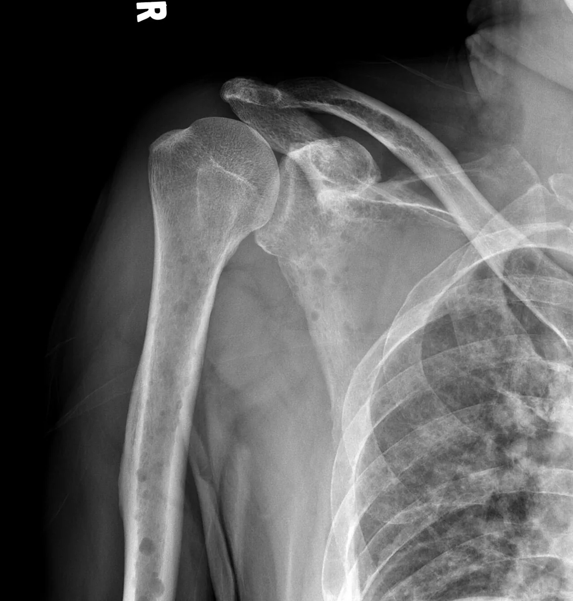
- Bone: myeloma cells secrete cytokines that activate osteoclasts and suppress osteoblasts; osteolytic lesions or diffuse osteoporosis result, preferentially in the pelvis, spine, ribs, femur, humerus, and skull.
- Kidney: most commonly deposition of light chains in the distal tubules (myeloma kidney) or hypercalcemia.
- Anemia due to kidney damage and suppression of erythropoiesis; susceptibility to infection due to lack of normal antibodies.
- AL amyloidosis in about 10 %, mostly with lambda light chains.
Clinical features
- Bone pain, persistent and often at night or at rest, especially in the back or thorax
- Pathologic fractures; vertebral collapse up to spinal cord compression with paraplegia
- Renal insufficiency, sometimes already at diagnosis
- Recurrent bacterial infections
- Symptoms of anemia, sometimes the only reason for evaluation
- Hypercalcemia: polydipsia, dehydration
- Peripheral neuropathy, carpal tunnel syndrome (especially with amyloidosis), and bleeding tendency are also common; hyperviscosity syndrome occurs only in a few
- Lymphadenopathy and hepatosplenomegaly are unusual.
Many myelomas are detected on routine tests, for example through elevated total protein, proteinuria, unexplained anemia, or renal insufficiency.
Diagnosis
Laboratory tests
- Complete blood count: anemia in about 80 %, usually normocytic and normochromic, with rouleaux formation on the smear; white cell and platelet counts usually normal.
- Serum chemistry: creatinine, urea, calcium (hypercalcemia at diagnosis in about 10 %), uric acid, LDH, beta-2 microglobulin, and albumin; sometimes a low anion gap.
Protein studies
- Serum protein electrophoresis: M-spike in about 80–90 %.
- Immunofixation of serum and concentrated 24-hour urine: determines the isotype and detects Bence Jones proteinuria.
- Serum free light chains (kappa, lambda, ratio): especially important in light chain myeloma; the ratio is also part of the SLiM criteria.
- Quantitative immunoglobulins (reduction of the uninvolved immunoglobulins).
Imaging and bone marrow
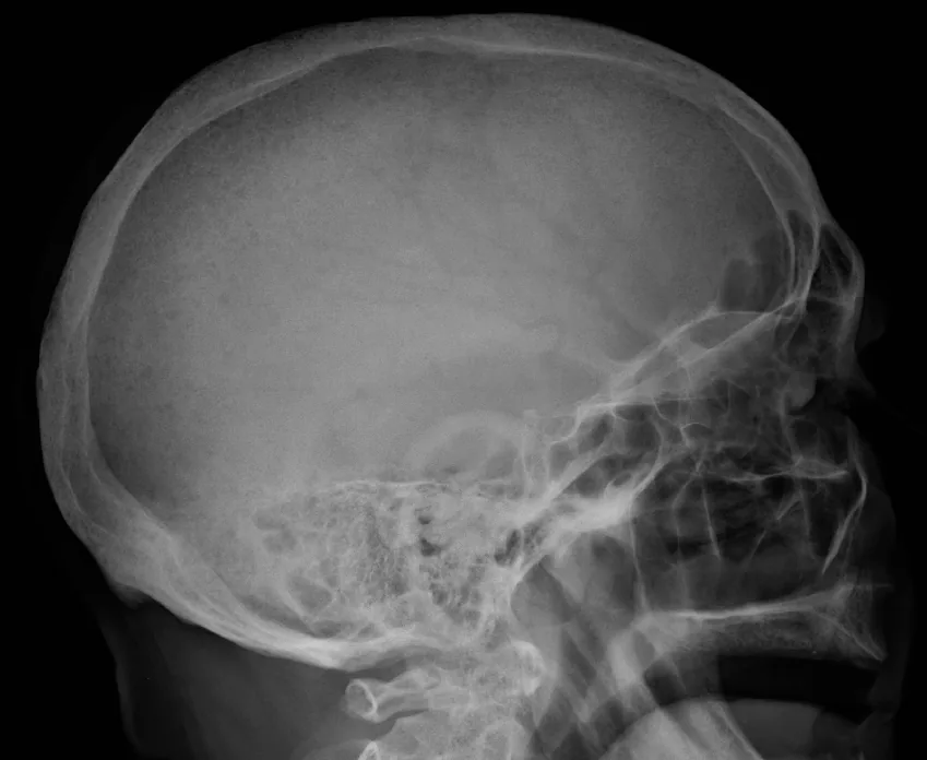
- Skeletal imaging: radiographic skeletal survey plus PET-CT or whole-body MRI, which detect bone lesions more sensitively; punched-out osteolytic lesions (e.g., in the skull) or diffuse osteoporosis in about 80 %. Bone scintigraphy is usually not helpful.
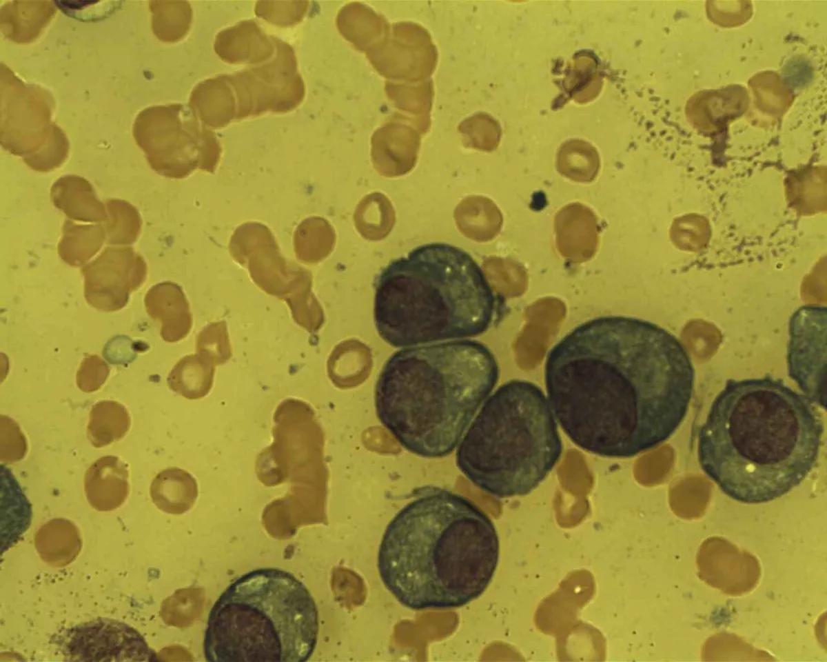
- Bone marrow aspiration and biopsy: sheets or clusters of plasma cells; because of patchy involvement, individual samples may show < 10 %. Flow cytometry, conventional cytogenetics, and FISH determine clonality and risk profile.
Keep learning in the app
Further reading (open access)
- MSD Manual Professional: Multiple Myeloma
- Clin Exp Med 2026: Monoclonal gammopathies, MGUS, SMM and multiple myeloma (PMC-Volltext)
- Fac Rev 2022: Monoclonal gammopathy of undetermined significance (PMC-Volltext)
- RKI, Zentrum für Krebsregisterdaten: Multiples Myelom
- Onkopedia-Leitlinie Multiples Myelom (DGHO)
Cross-references
More topics: Haematology & oncology
- Anemia (classification and work-up)
- Hemolytic anemias
- Acute myeloid leukemia (AML)
- Iron deficiency anemia
- Non-Hodgkin lymphomas
- Vitamin B12 deficiency and pernicious anemia
- Paraneoplastic syndromes
- Autoimmune hemolytic anemia (AIHA)
- Febrile neutropenia
- Immune thrombocytopenia (ITP)
- Renal anemia
- Aplastic anemia
Note: Learning content for medical education – not a treatment recommendation and no substitute for diagnosis or treatment decisions in individual cases. Treatment and management are deliberately not covered on this page.