Aplastic anemia
Board exam relevance: in 4 of 105 exam reports · rank 90- Synonyms
- bone marrow failure, panmyelopathy, marrow aplasia, severe aplastic anaemia
- Specialty
- Internal medicine · Haematology & oncology
- Images
- Histology 1
- Last updated
- 10/2026 · Dr. Pascal Bafteh
Contents
Images (1)
Definition
Aplastic anemia (panmyelopathy) is a disorder of the hematopoietic stem cell with hypoplasia or aplasia of the bone marrow and a reduction of at least two cell lines in the blood (bicytopenia or pancytopenia). The marrow is hypocellular and fatty, but neither infiltrated by tumor cells nor fibrotic.
Marrow damage due to ionising radiation or obligatorily myelotoxic substances is not included. Isolated aplasia of the red cell line (pure red cell aplasia) is a separate entity.
Classification
Severity (two of three blood criteria)
- Non-severe (moderate) aplastic anemia: neutrophils below 1.2 G/l, platelets below 70 G/l, reticulocytes below 60 G/l; plus hypocellular marrow
- Severe aplastic anemia: neutrophils below 0.5 G/l, platelets below 20 G/l, reticulocytes below 60 G/l; plus marrow cellularity below 25 % (or 25–50 % with less than 30 % hematopoietic cells)
- Very severe aplastic anemia: as for the severe form, with neutrophils below 0.2 G/l as an obligatory criterion
Occurrence & epidemiology
The incidence in central Europe is about 2–3 cases per million inhabitants per year. The acquired form can occur at any age; the age distribution shows two peaks, between 10 and 25 years and over 60 years. There is no sex predilection.
Aetiopathogenesis
In most acquired cases the immune system, mainly via T lymphocytes, attacks the hematopoietic stem cells; often no trigger can be found (idiopathic form). Known triggers and causes:
- Drug-induced as an idiosyncratic reaction, e.g. by certain anti-inflammatory agents, anticonvulsants, antithyroid drugs, antimalarials and anti-infectives
- Chemicals and toxins: benzene, inorganic arsenic, insecticides
- Infections: hepatitis without evidence of known hepatitis viruses, Epstein-Barr virus, cytomegalovirus
- Pregnancy
- Inherited bone marrow failure syndromes, which may present only in adulthood (“cryptic”): Fanconi anemia, dyskeratosis congenita and other telomeropathies, Shwachman-Diamond syndrome, GATA2 deficiency
Many patients also have a PNH clone (AA/PNH syndrome) or clonal hematopoiesis; there is a risk of progression to myelodysplasia or acute myeloid leukemia.
Clinical features
Onset is usually insidious over weeks to months, occasionally acute. Symptoms result from the cytopenia:
- Anemia: weakness, easy fatigability, exertional dyspnea
- Thrombocytopenia: petechiae, ecchymoses, gingival and conjunctival bleeding
- Neutropenia: severe infections, e.g. oral and pharyngeal ulcers, necrotising gingivitis or tonsillitis, pneumonia, cellulitis
Lymphadenopathy, hepatomegaly or splenomegaly argue against aplastic anemia. Clues to inherited forms are skin pigment abnormalities, oral leukoplakia, nail dystrophy, pulmonary fibrosis or liver cirrhosis and malformations.
Histology
Blood and bone marrow
- Blood smear: unremarkable red cell morphology, normocytic to slightly macrocytic; leukopenia due to granulocytopenia and monocytopenia without immature precursors; no giant platelets
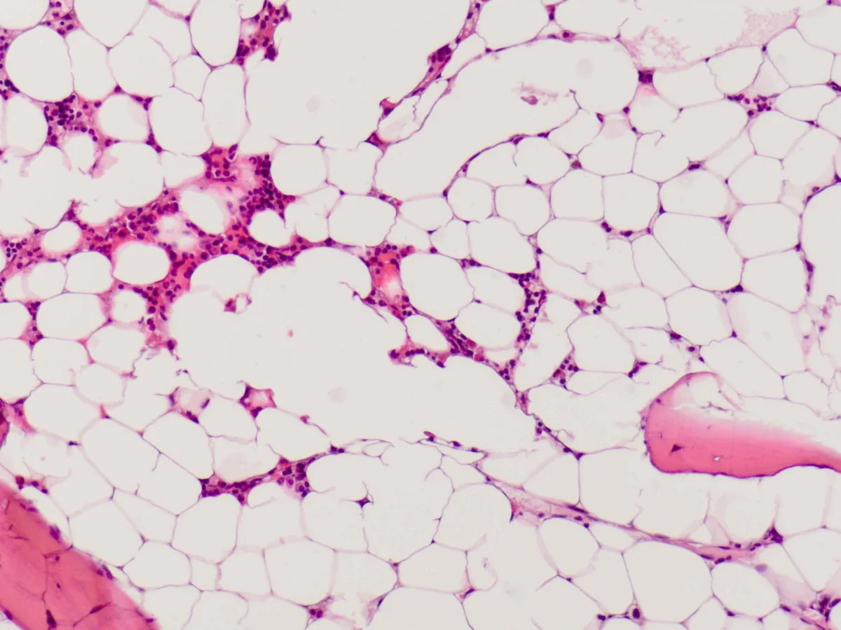
- Bone marrow biopsy: hypoplasia to aplasia with fatty marrow, often patchy with erythropoietic “hot spots”; increased T lymphocytes without clonality; no tumor cell infiltration, no fibrosis
Diagnosis
- Blood count with reticulocytes: bicytopenia or usually tricytopenia, reticulocytopenia or an inadequate reticulocyte production index
- Bone marrow aspiration and biopsy (both required, biopsy length at least 15 mm) with cytogenetics (karyotype, FISH) and molecular testing to distinguish myelodysplasia and leukemia
- Flow cytometry for a PNH clone; a PNH clone argues against an inherited bone marrow failure syndrome
- Search for inherited forms, especially in young people, with a positive family history or malformations: chromosome breakage test (Fanconi anemia), telomere length measurement, genetic sequencing
- Search for causes: drug and exposure history, hepatitis and viral serology (EBV, CMV)
Keep learning in the app
Further reading (open access)
Cross-references
More topics: Haematology & oncology
- Anemia (classification and work-up)
- Hemolytic anemias
- Acute myeloid leukemia (AML)
- Iron deficiency anemia
- Non-Hodgkin lymphomas
- Vitamin B12 deficiency and pernicious anemia
- Multiple myeloma
- Paraneoplastic syndromes
- Autoimmune hemolytic anemia (AIHA)
- Febrile neutropenia
- Immune thrombocytopenia (ITP)
- Renal anemia
Note: Learning content for medical education – not a treatment recommendation and no substitute for diagnosis or treatment decisions in individual cases. Treatment and management are deliberately not covered on this page.