Immune thrombocytopenia (ITP)
Board exam relevance: in 5 of 105 exam reports · rank 69- Synonyms
- idiopathic thrombocytopenic purpura, autoimmune thrombocytopenia, Werlhof disease, low platelets, immune thrombocytopenic purpura
- Specialty
- Internal medicine · Haematology & oncology
- Images
- Clinical 2
- Last updated
- 10/2026 · Dr. Pascal Bafteh
Contents
Images (2)
Definition
Immune thrombocytopenia (ITP), formerly Werlhof disease or idiopathic thrombocytopenic purpura, is an acquired, non-hereditary autoimmune disorder with isolated thrombocytopenia below 100 G/l. Anemia and leukopenia are typically absent (except for iron deficiency anemia due to bleeding). ITP is a diagnosis of exclusion: there is no test that proves it.
Primary ITP is distinguished from secondary forms associated with underlying conditions such as other autoimmune diseases, lymphoproliferative disorders, immunodeficiency or infections (e.g. HIV, hepatitis C).
Classification
Phases by disease duration
- Newly diagnosed: up to 3 months after diagnosis; spontaneous normalisation is still common
- Persistent: 3–12 months after diagnosis
- Chronic: longer than 12 months; spontaneous normalisation now becomes unlikely
Occurrence & epidemiology
In adults, the incidence is 0.2–0.4 new cases per 10,000 inhabitants per year and the prevalence 0.9–2.6 per 10,000; in children and adolescents the incidence is 0.2–0.7 and the prevalence 0.4–0.5 per 10,000. In children ITP often occurs acutely after an infection and is usually self-limiting, whereas in adults it becomes chronic in about 60–70 %. About 8 % of people with common variable immunodeficiency (CVID) also have ITP.
Aetiopathogenesis
Autoantibodies, usually against platelet surface glycoproteins (e.g. GP IIb/IIIa), lead via Fc receptors to increased platelet destruction in the spleen and liver. Further mechanisms are complement damage, desialylation with hepatic clearance, direct damage by autoreactive T lymphocytes and inhibition of platelet production: autoantibodies damage megakaryocytes, and thrombopoietin production is relatively reduced.
In children, antibody production is often triggered by viral infections; in adults the trigger usually remains unknown. In some countries (e.g. Japan, Italy) there is an association with Helicobacter pylori.
Clinical features
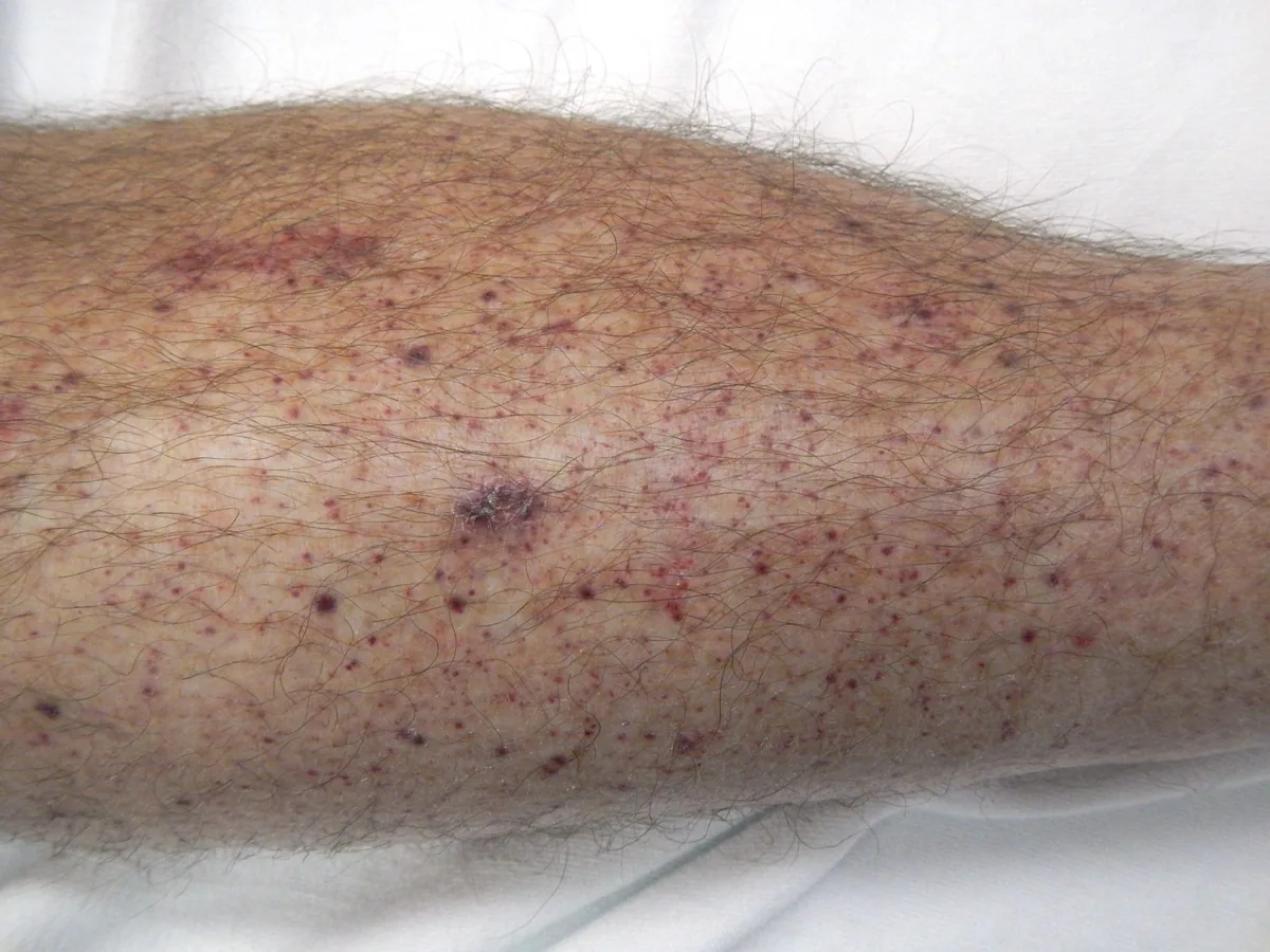
Many patients are asymptomatic and detected by a routine blood count; at diagnosis about 20–30 % of adults have no bleeding signs. The platelet-type bleeding pattern is typical:
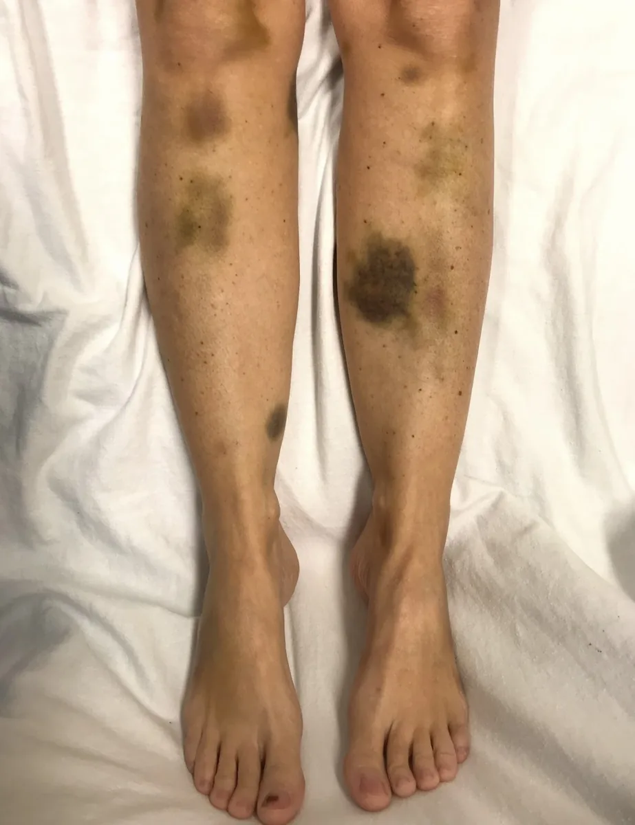
- non-palpable petechiae, especially on the legs; purpura and ecchymoses
- mucosal bleeding in the mouth and nose, heavy menstrual bleeding, urogenital bleeding
- increased bleeding and bruising after minor injuries
- rarely severe internal bleeding, intracerebral hemorrhage in less than 1–2 %
- fatigue, cognitive impairment
- increased thrombotic risk despite thrombocytopenia
The spleen is not enlarged, except with a coexisting viral infection or autoimmune hemolytic anemia (Evans syndrome). Palpable purpura suggests vasculitis, while extensive hematomas and joint bleeds suggest a coagulation factor disorder.
Histology
Blood smear and bone marrow
The smear shows isolated thrombocytopenia with large platelets and otherwise normal red and white cell morphology; no schistocytes. The bone marrow shows normal or increased megakaryocytes with otherwise unremarkable hematopoiesis.
Diagnosis
- Blood count in EDTA and citrate blood to exclude pseudothrombocytopenia (EDTA-induced platelet clumping)
- Blood smear reviewed by an experienced hematologist: platelet size and granularity, exclusion of TTP (schistocytes), inherited thrombocytopenias and leukemia
- A raised mean platelet volume and raised immature platelet fraction (with counts below 50 G/l) are consistent with ITP
- Coagulation (prothrombin time/INR, aPTT, fibrinogen) and liver tests
- History: drugs, alcohol, infections, pregnancy, previous thrombosis, family history
- Serology for HIV and hepatitis C, testing for Helicobacter pylori; immunoglobulins to exclude immunodeficiency; direct Coombs test with coexisting anemia (Evans syndrome)
- Platelet autoantibodies only in persistent or chronic ITP or an atypical course (only a positive result is informative)
- Bone marrow: with atypical findings, other blood count abnormalities, at older age (from about 60 years) or with an atypical course
Keep learning in the app
Further reading (open access)
Cross-references
More topics: Haematology & oncology
Note: Learning content for medical education – not a treatment recommendation and no substitute for diagnosis or treatment decisions in individual cases. Treatment and management are deliberately not covered on this page.