Acute myeloid leukemia (AML)
Board exam relevance: in 13 of 105 exam reports · rank 17- Synonyms
- acute myelogenous leukemia, acute myelocytic leukemia, blood cancer, acute leukemia, APL
- Specialty
- Internal medicine · Haematology & oncology
- Images
- Blood smear & cytology 4 · Histology 1
- Last updated
- 10/2026 · Dr. Pascal Bafteh
Contents
Images (5)
 Blood smear & cytology
 Blood smear & cytology
 Blood smear & cytology
 Blood smear & cytology
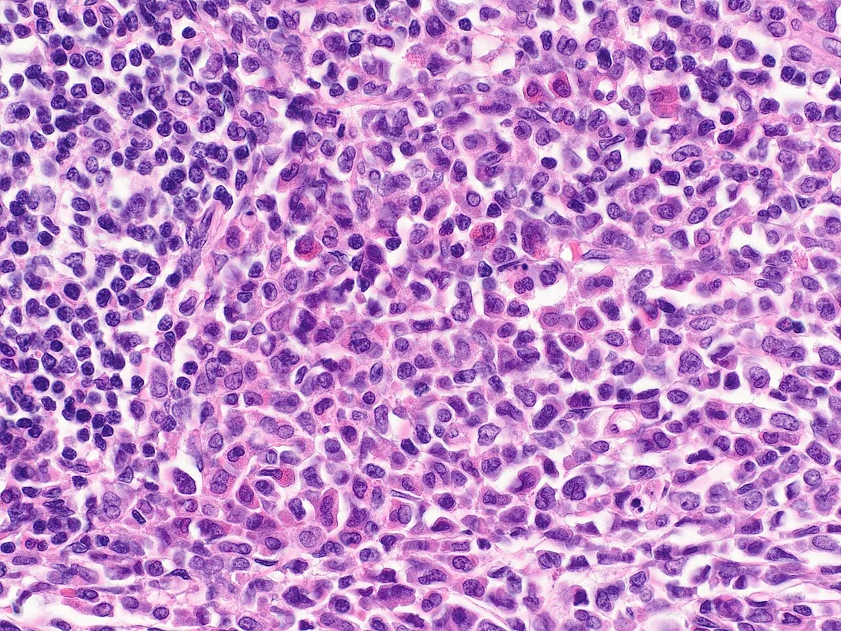 Histology
Definition
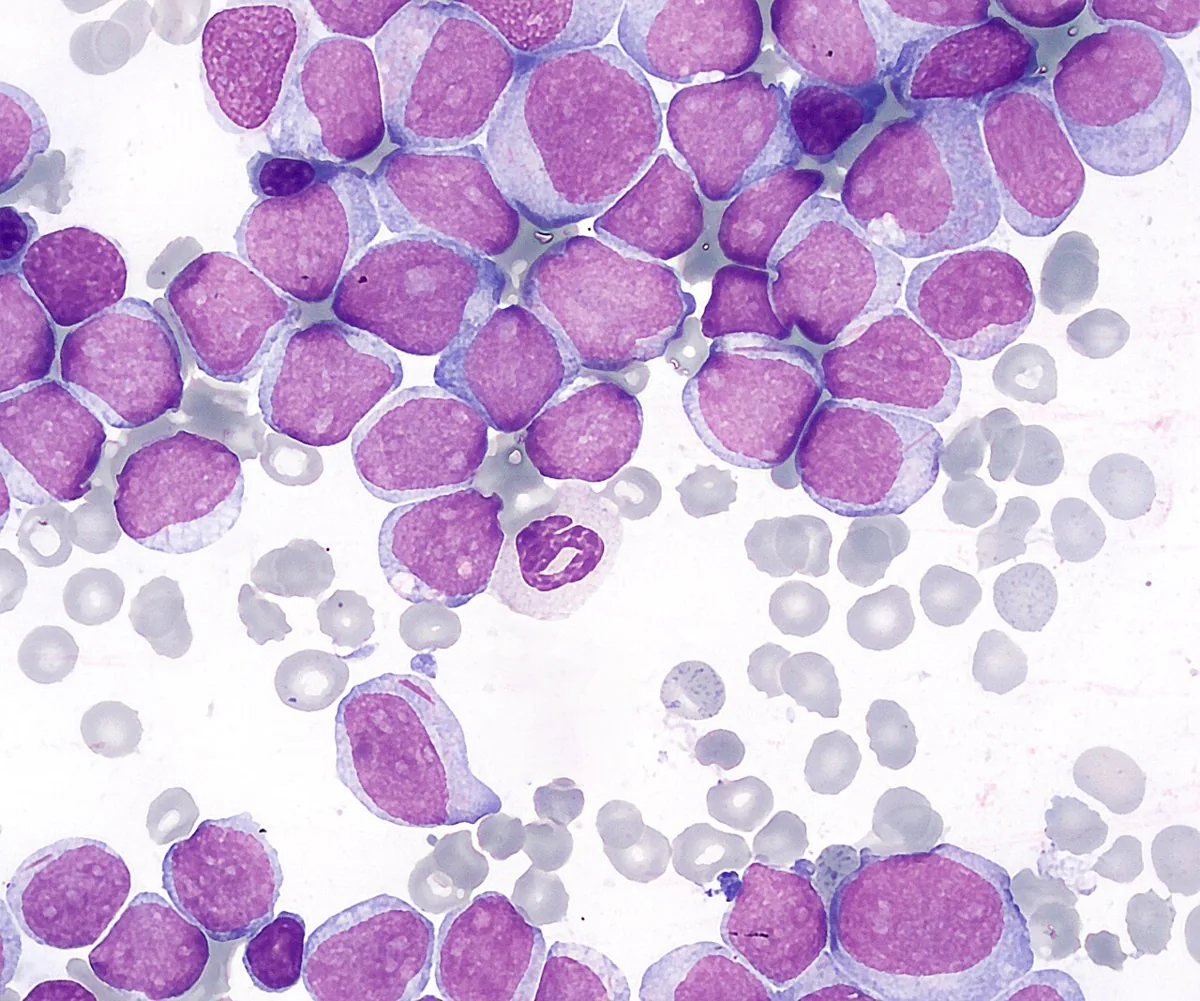
Acute myeloid leukemia (AML) is a malignant disease of myeloid progenitor cells. An acquired series of genetic aberrations, usually at the level of the pluripotent stem cell, leads to uncontrolled proliferation, disturbed maturation, and reduced apoptosis. Immature blasts displace normal hematopoiesis in the bone marrow and may appear in the blood. The WHO diagnostic threshold is 20 % blasts in blood or bone marrow; for certain defining genetic abnormalities, a lower blast count is sufficient.
Classification
The 2022 WHO classification separates AML with defining genetic abnormalities from AML defined by differentiation. The International Consensus Classification (ICC) follows a similar, likewise genetics-based structure but sets different blast thresholds: most genetically defined types require ≥ 10 % blasts, and 10–19 % blasts without an AML-defining abnormality are termed MDS/AML. The following list reflects the WHO types.
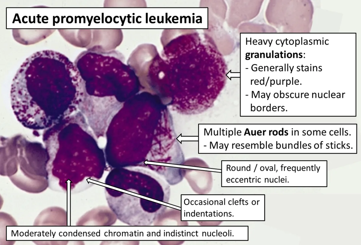
- Acute promyelocytic leukemia (APL) with PML::RARA fusion, t(15;17): 10–15 % of all AML, younger patients (median age 31 years), frequently with a coagulation disorder up to disseminated intravascular coagulation (DIC)
- AML with RUNX1::RUNX1T1, t(8;21), and AML with CBFB::MYH11, inv(16) or t(16;16)
- AML with KMT2A, MECOM, or NUP98 rearrangement
- AML with NPM1 mutation: irrespective of the blast count
- AML with CEBPA mutation and AML with BCR::ABL1: only these two genetically defined types require ≥ 20 % blasts
- AML, myelodysplasia-related (AML-MR): ≥ 20 % blasts with MDS-typical cytogenetic or molecular abnormalities, de novo or following MDS or MDS/MPN
- AML after prior cytotoxic exposure: usually 1–10 years after alkylating agents or topoisomerase II poisons
- Myeloid sarcoma: extramedullary infiltration by myeloblasts, e.g., of the skin (leukemia cutis) or the gingiva
Occurrence & epidemiology
AML is predominantly a disease of adulthood; the median age at diagnosis is 68 years. Men are affected slightly more often than women. The lifetime risk in the United States is about 0.5 %. According to RKI data, about 13,450 people in Germany were diagnosed with leukemia in 2022; AML is, together with CLL, among the most common forms.
Aetiopathogenesis
The cause is a series of acquired genetic aberrations in a hematopoietic stem or progenitor cell. Known risk factors:
- ionizing radiation and benzene
- prior cytotoxic drugs (alkylating agents, topoisomerase II poisons)
- preexisting myeloid neoplasms (MDS, myeloproliferative neoplasms)
- congenital syndromes such as Down syndrome (trisomy 21), Fanconi anemia, Bloom syndrome, ataxia-telangiectasia, or Li-Fraumeni syndrome
Alkylating agents typically cause deletions and unbalanced translocations, topoisomerase II poisons balanced translocations, particularly KMT2A rearrangements.
Clinical features
Leading symptoms
Symptoms are often present for only days to weeks before diagnosis. They arise mainly from displaced hematopoiesis:
- Anemia: fatigue, weakness, pallor, exertional dyspnea, tachycardia, exertional chest pain
- Thrombocytopenia: mucosal bleeding, bruising, petechiae and purpura, nose and gum bleeding, heavy menstrual bleeding; less often spontaneous intracranial or intra-abdominal hemorrhage
- Granulocytopenia: fever, severe or recurrent bacterial, fungal, and viral infections
Organ infiltration and complications
- Organ infiltration is less common in AML than in ALL; the liver, spleen, and lymph nodes may be enlarged.
- Bone marrow and periosteal infiltration cause bone and joint pain.
- Leukemia cutis: papules, nodules, or plaques, erythematous, brown, hemorrhagic, or violaceous; extramedullary infiltrates affect only about 5 % of patients overall.
- Meningeal infiltration: cranial nerve palsies, headache, visual or auditory symptoms, altered mental status.
- APL: coagulation disorder with DIC and bleeding tendency already at diagnosis.
Diagnosis
Blood count and smear
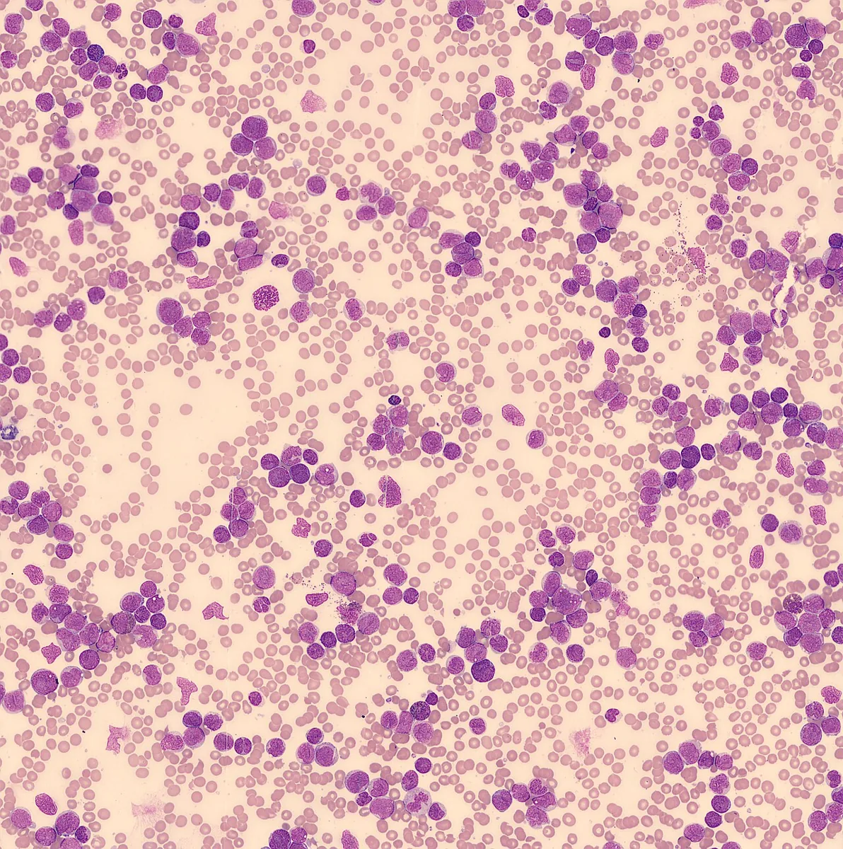
- Pancytopenia with peripheral blasts suggests acute leukemia; blasts may account for up to 90 % of white cells.
- The white cell count may be increased, normal, or decreased; severe anemia and thrombocytopenia are the rule at diagnosis.
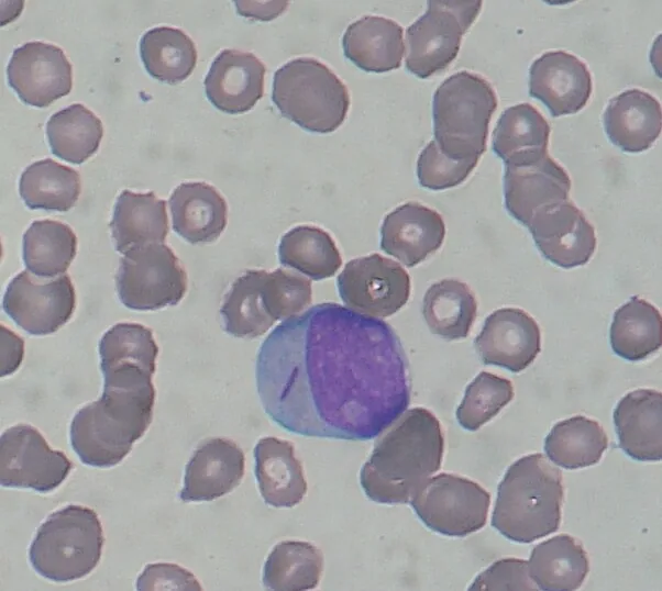
- Auer rods (rod-shaped azurophilic inclusions formed from crystallized myeloperoxidase-rich granules) in the cytoplasm of myeloblasts indicate AML.
- A leukemoid reaction in infection never shows high blast counts.
Bone marrow, immunophenotype, and genetics
- Bone marrow aspiration and biopsy are routine; the blast count in AML is typically between 25 and 95 %.
- Cytochemistry: myeloperoxidase staining positive in cells of myeloid origin.
- Immunophenotyping: myeloid markers such as CD13, CD33, CD34, and CD117; distinguishes AML from ALL.
- Cytogenetics and molecular genetics: determine the genetic AML type (e.g., PML::RARA, RUNX1::RUNX1T1, CBFB::MYH11, NPM1, CEBPA, TP53, KMT2A).
Additional tests
- Coagulation tests in patients with bleeding, especially when APL with DIC is suspected
- Cultures in patients with signs of infection
- Serum chemistry: transaminases, creatinine; falsely low glucose (pseudohypoglycemia) with high cell counts
- CT of the head in patients with neurologic symptoms
- Echocardiography to document baseline cardiac function
Keep learning in the app
Further reading (open access)
- MSD Manual Professional: Acute Myeloid Leukemia (AML)
- Khoury et al., Leukemia 2022: WHO-Klassifikation (5. Aufl.), myeloische Neoplasien (PMC-Volltext)
- MSD Manual Professional: Overview of Leukemia
- RKI, Zentrum für Krebsregisterdaten: Leukämien
- Arber et al., Blood 2022: International Consensus Classification (ICC) myeloischer Neoplasien und akuter Leukämien (PMC-Volltext)
Cross-references
More topics: Haematology & oncology
Note: Learning content for medical education – not a treatment recommendation and no substitute for diagnosis or treatment decisions in individual cases. Treatment and management are deliberately not covered on this page.